7
Facial Rejuvenation with Autologous Fat Injections
Natural-appearing rejuvenation should be one of the overall goals of facial aesthetic surgery. For many years, the focus of surgical procedures has been on lifting lax skin, fat, and muscle. The face ages not only with ptosis, however, but also with fat atrophy. To correct these atrophic changes, filling materials are used. Although the search for the perfect filler material continues, fat is clearly coming to the forefront as a safe, noninflammatory, autologous material that plays a significant role in facial rejuvenation. This chapter reviews the history of fat transfer, our approach to fat transfer, and the controversies surrounding fat transfer to provide the cosmetic surgeon with a comprehensive review of this helpful technique of rejuvenation.
Neuber, who transplanted fat grafts from the arm to correct a facial soft tissue defect in 1893, presented the initial concept of autologous fat transfer.1 He noted some success to transferring en bloc fat grafts to sites as long as they were smaller than an almond in size. Advances have been refined over the years from transferring bulk parcels of excised fat tissue from one site to another site to transferring liposuctioned fat strands to precise locations in 1 to 2 mL amounts. The history of fat transfer is reviewed excellently in many articles.2–7 Bruning implemented fat injection to correct postrhinoplasty defects.8 Peer helped to determine that ∼50% of fat remained present after fat transplant.9 In 1976, Fischer and Fischer performed fat extraction with a cellusuctiotome.10 More recent historical contributions in fat transfer to the skin come from Illouz, who refined the technique of liposuction and reinjected fat from liposuction procedures to correct surgical soft tissue defects.11,12 Fournier also made a key contribution by demonstrating “microlipoinjection” fat harvesting with 13-gauge needles attached to syringes combined with subcutaneous injections.13 With the innovation of Klein’s introduction of tumescent anesthesia,14 infiltration of fat with large amounts of dilute anesthesia facilitated fat harvesting. Many authors have subsequently made key refinements in fat harvesting and injection techniques and optimization to promote fat injection survivability.4,15–20
In addition to facial rejuvenation, fat transfer has been used to solve the problems of unequal distribution of fat in facial hemiatrophy and lipodystrophy.21 Many specialties including ophthalmology, neurosurgery, orthopedics, otolaryngology, and urology have used fat transfer techniques as a resource to restore structure and function to their respective areas of practice.22–24
There are many different techniques of fat transfer and supportive theories for these techniques. This has led to some confusion for many in regard to this procedure. Techniques range from the injection of only fresh fat, to the injection of only frozen fat, or a combination of both. Different authors also disagree on fat injection techniques, including fat preparation, instrumentation, and the method by which it is injected. Due to these wide variables, it is often difficult to compare different results in the literature. In the technique described here, fresh fat is injected on the initial visit, and frozen fat is injected on follow-up appointments.
To understand where to enhance the aging face with fat injection, one must first understand typical aging patterns of the face. Wrinkles, laxity, ptosis, fat atrophy, and fat hypertrophy tend to occur over time. Wrinkles, dyschromia, and telangiectasia are well treated with various procedures including ablative and nonablative laser treatments and botulinum toxin injections. Laxity and ptosis, which are usually related to intrinsic aging, manifest as brow ptosis, malar ptosis, jowl formation, and neck aging. These have typically been corrected with procedures including brow lifts, face and neck lifts, blepharoplasty, and treatments with a nonablative radiofrequency device (ThermaCool, Thermage Inc., Hayward, California). Although these procedures are adept at controlling laxity, patients often complain that they appear drawn or gaunt. The patient loses a perceived fullness in the face after these areas are lifted. After the lift, the remaining fat atrophy is more evident with hollowness in the buccal fat area, nasolabial or melolabial folds, mental area, lips, temporal fossa, and periorbital tissues. The lower two thirds of the face require volume replacement to restore the roundness and softness of the originally younger face. Volume can also be used in the upper face to create a more youthful contour of the forehead and ocular areas. Occasionally, areas of fat hypertrophy, which tend to occur in the submental region, the infraorbital fat pads, and jowling along the jawline, may require fat removal to re-create the soft fluid lines of a younger face. The progression of facial aging may be delayed through the usage of fat as a filler to structurally augment the supporting framework. The option of rejuvenation through fat injection is especially helpful for the younger generation, which may not need the lifting correction of a conventional surgical facelift. Thus fat injections are used in both younger and older patients to restore a youthful, fuller facial appearance.
Many of these indications can be corrected with various filler substances. When evaluating a filler substance, one wants to look for several characteristics. The substance ideally provides persistent and predictable correction via a reproducible technique. It is easily obtainable or fabricated, noninflammatory, noncarcinogenic, nonmigratory, multipurpose, biodegradable, and cost effective. There are quite a few available filler substances to restore the face, and many of them meet, to varying degrees, the properties of the ideal filler substance. However, fat is the only autologous long-lasting, noninflammatory product available. Although controversy exists as to the length that fat lasts, it is still an outstanding filler substance. The main limitations to fat injections are the inability to inject fat into small, superficial rhytids as well as a lack of donor site fat in certain individuals.
 Indications for Lipotransfer
Indications for autologous fat transfer include aging-related changes of the face, scars, and nonfacial changes. When approaching fat as a filler substance, it can be used to correct the nasolabial folds and oral commissures. It can also be fanned into areas to correct atrophy of the chin, buccal fat loss, and temporal wasting. Fat can also be placed into the infrabrow area to re-create upper lid fullness, which is associated with the perception of youth. Fat may also be used in the infraorbital area and nasojugal groove, but with caution, because these areas have a tendency to resorb unevenly, leaving small bumps. Correction of malar pouches can be accomplished by using fat to fill this area, as well as laterally to augment the cheekbones. Fat is also helpful for lip augmentation. Scarring indications include distensible acne scarring, trauma-related scarring, and atrophic congenital or acquired defects. Other enhanceable areas include the hands,25 and other body contour defects.
 Patient Selection and Preoperative Evaluation
Any patient with the appropriate indications and sufficient donor fat can be a good candidate for this surgical procedure. Because this particular technique is performed with local anesthesia, an ideal candidate should not have extreme concerns in regard to being awake. In such instances, this procedure can be performed with intravenous sedation. Preoperative evaluation includes the benefits of the procedure for the patient, as well as a detailed discussion of the inherent risks. It is important that patients understand the filling nature of this procedure, and the slow building process that ensues. Routine preoperative laboratories and other indicated tests and medical clearance are performed. Antibiotics are started on the morning of the procedure and are continued for 5 days.
 Lipoharvesting
These authors prefer to use either outer thigh or hip lipocytes because these areas are usually the easiest to obtain and thus less traumatic for the patient. The authors use abdominal fat as a second choice because this fat tends to be more fibrous and, thus, more difficult to reinject. Overall, the site is dictated by where a sufficient quantity of donor material is obtainable. The selected site is demarcated with a surgical pen while the patient is standing to ensure a smooth contour. Most patients appreciate preoperative relaxation with oral valium dosages of 5 to 10 mg and/or one oral Vicodin.
Fat harvesting is accomplished while the patient is recumbent with the maintenance of sterile technique in an accredited surgical suite. After the patient is prepped and draped, a small wheal of 1% lidocaine is injected at the sites of insertion of the cannula. Approximately 120 mL of the tumescent solution (Table 7-1) are injected into each donor site utilizing 60 mL syringes and a Luer locking syringe 20 cm, 2 mm radial injection cannula. The tumescent anesthesia is injected with a fanning radial motion into the donor site. Within 15 minutes, the tumescent solution has distributed to provide adequate anesthesia and vasoconstriction to minimize bleeding. Harvesting is performed with a blunt-tipped triport aperture microcannula 17 cm in length with a 2 or 3 mm aperture attached to a 60 mL syringe with a snapper device to maintain suction. A fanning radial motion with the dominant hand is used to remove 35 to 60 mL of fat cells per site (Fig. 7-1.). The syringe is capped and gravity is used to separate the fat cells, the supernatant, from the serosanguinous infranatant and intervening triglyceride layer. The infranatant and triglyceride layers are then discarded. If there are large quantities of heme, the remaining fat is washed with sterile saline and allowed to separate with gravity again. No centrifugation is performed. This supernatant fat is transferred via female-female transfer Luer locks to 3 mL syringes labeled with the patient’s name, Social Security number, and procedure date. The harvested fat is stored in a centrally alarmed freezer at -20°C for 12 to 18 months.
| 225 mL | Intravenous normal saline |
| 12.5 mL | 2% lidocaine (250 mg) |
| 1 mL | Adrenaline 1:1000 |
 Lipoinjection Technique
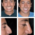
The areas to be injected for facial rejuvenation are reviewed and marked as shown (Fig. 7-2.). For the initial injection of the freshly harvested tissue, either or both local and regional anesthetic blocks are performed. The 3 mL syringe is attached to an 18-gauge, 2-inch, blunttipped, side-port microcannula. An 18-gauge needle is used to make initial puncture sites. The microcannula is placed into the deep subcutaneous plane parallel to the surface of the skin (Fig. 7-3.). This placement may be either into the muscle tissue or into the subcutaneous fat layer. The fat is then injected on slow withdrawal in a retrograde fashion. The cannula is repositioned, either in a more superficial plane as in the nasolabial folds, or more laterally as in the cheek and chin area, and more fat is injected. Gentle compression is used to mold the fat (Fig. 7-4.). Enough fat is injected to achieve a 15 to 35% overcorrection. Injections into the lip can be into the vermilion border to define the lip or into the body of the lip to augment the size. Ice packs are applied to minimize swelling and discomfort.
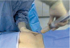
Figure 7-1 Lipoharvesting technique. A fanning radial motion with a 60 mL syringe with snapper device removes 35 to 60 mL of fat cells from the outer thigh.
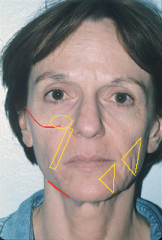
Figure 7-2 Injection areas. The cheek, nasolabial, malar cheek, and chin areas outlined in yellow are injected with autologous fat to provide filling and fullness in those areas. The cheekbone and jaw areas outlined in red are injected to provide structural augmentation.
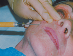
Figure 7-3 Lipoinjection technique. After local anesthesia or regional blocks (lips), and passage of fresh fat once through a Luer-Luer transfer adapter, fat is injected into the nasolabial folds through 18-gauge needle puncture sites via an 18-gauge, 2-inch, blunt-tipped, side-port microcannula.
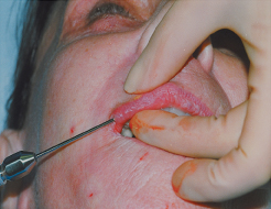
Figure 7-4 Lipoinjection technique. The microcannula is placed into the deep subcutaneous plane parallel to the surface of the skin, and the fat is injected on slow withdrawal in a retrograde fashion. The cannula may be repositioned in more superficial planes, and additional injections may be performed. Gentle compression with the fingers is used to mold the injected fat. With fresh fat, the goal is a 15 to 35% overcorrection.
 Follow-up Injections of Frozen Fat
Follow-up injections of frozen fat are repeated at 4- to 6-week intervals. To avoid specimen error, the patient’s name and Social Security number are checked and initialed by the physician and assistant. The patient thaws the syringe of fat under the axilla or hands. Any small bit of watery infranatant is decanted. The fat is passed through a Luer-Luer transfer adapter and the syringe is attached to a 20-gauge, 1.5-inch needle for injection. Typically, no anesthesia is used except for blocks when the lips are injected. The frozen fat is injected in a similar manner to fresh fat lipoinjection already explained. No overcorrection is obtained, however. Ice packs are applied to decrease swelling and discomfort.
 Adverse Sequelae and Complications
Related posts:
Stay updated, free articles. Join our Telegram channel

Full access? Get Clinical Tree


