Acknowledgments
To our team, our families, and our patients.
Introduction
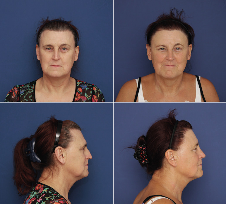
From a very early age, everyone is able to recognize whether a face is male or female, long before learning about genital differences. The perception of gender through facial features occurs in a single glance, involuntarily, and definitively. While other parts of the body can be hidden, camouflaged, or exaggerated to appear more feminine, it is difficult to create female facial features without the appropriate surgical reassignment of facial gender. It is for this reason that an individual in the process of transitioning from man to woman may want this surgery in order to modify their face and better integrate into society, the workplace, and the family. The surgical reassignment of facial gender is generating considerable interest among trans women and trans-health professionals. Modifying facial gender within the transition protocol is without doubt as important as hormone therapy and genital reconstruction.
Definition and Differences
From a technical point of view, Facial Feminization Surgery (FFS) can be defined as the set of surgical procedures associated with different surgical specialties (Oral and Maxillofacial Surgery, Craniofacial Surgery, Plastic and Reconstructive Surgery) designed to soften and modify facial features perceived as masculine, exaggerated, or nonharmonic, and which, therefore, are decisive in the visual identification of facial gender. On a somewhat more philosophical note, a much more specific term would be more appropriate for the type of surgery discussed in this chapter: Facial Gender Confirmation Surgery (FGCS). The popularly known concept of FFS is, in fact, so broad that it can include groups of patients without any gender dysphoria symptoms, for whom the techniques included in FFS are absolutely indicated (e.g., cis females with unusually prominent supraorbital ridge or cis males with especially wide jaws). This chapter focuses exclusively on male-to-female FGCS (MtF FGCS) due to the low incidence of and indication for surgical treatment in female-to-male patients and its near absence in the scientific literature reviewed.
When evaluating, diagnosing, and planning a patient’s feminization needs, it is essential to understand the differences between male and female facial features. Generally speaking, the male facial skeleton has some well-defined features that distinguish it from its female counterpart. The basic pillars for the visual identification of facial gender are the frontonasoorbital complex, the nose, and the maxillomandibular complex. Other aspects, structural and not, can also influence this identification, such as thyroid cartilage (Adam’s apple), hairline format, cheekbones, the upper lip, facial hair, skin type and quality, and the distribution of facial fat.
Gender differences: primary aspects
Genetic sex is determined at conception, but gonadal hormones play a vital role in the differentiation of male and female phenotypes throughout human development. Prenatal and adolescent levels of testosterone, the most abundant androgen, condition the appearance of facial features related to gender identity, which can be divided into primary aspects (structural) and secondary aspects (hormone–dependent).
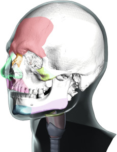
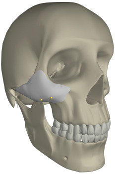
These differentiating features appear in the frontonasoorbital complex, the nose, the malar region, the upper lip, the jaw and chin complex, and the thyroid cartilage ( Fig. 8.1 ). The development of these structures under hormonal influence is not reversible, and thus these features, which determine a significant part of an individual’s facial gender, can only be approached and modified using surgery, always respecting the intrinsic architecture and anatomy of the craniofacial skeleton.
Frontonasoorbital Complex
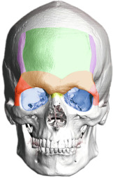
This area is quite possibly the greatest determinant of facial gender. The region encompasses the forehead surface, the supraorbital ridge (frontal bossing), the eye sockets, the frontomalar buttresses, the temporal ridges, and the frontonasal transition ( Fig. 8.2 ). The supraorbital ridge is almost invariably much more strongly developed in the male than in the female, although, typically, all of these areas are more pronounced and have greater bone volume in the male skeleton than in the female skeleton. The forehead contour in the female is higher, smoother, more vertical, and may be rounded to the point of forward protrusion. It determines the position of the eyebrows and the positioning of the periorbital soft tissues like the eyelids.
Nose
From the perspective of gender difference, the male nose is usually larger than the female because it has a greater component of bone and cartilage. The male nasal bones are larger and tend to meet in the midline at a sharper angle. Female noses tend to be narrower, the tip is often sharper, and the nostrils may be smaller. The frontonasal transition can be another important area with regard to facial gender differences. In males, the angle formed by the transition between forehead and nose tends to be more acute. However, the nose has characteristics conditioned by ethnicity and age that are almost as important as gender-based differences.
Malar Region
The cheek area (zygomatic-malar region of the facial skeleton) usually has some structural differences that must be defined, since it can readily lead to confusion with regard to facial feminization. As a general rule, the malar bone volume is greater in men, which can result in well-defined cheeks. However, prominent round cheeks in the middle third of the face are compatible with femininity, due to a greater concentration of fat in this area in women (e.g., the greater volume is due not to the bone but to the soft tissues). This has specific implications when it comes to deciding the best treatment in this region.
Upper Lip
On the whole, the distance between the upper lip and the nose (cutaneous portion) is greater in men than in women. Additionally, a recent study done by Penna et al. found that the ratio of the upper vermillion height/mouth-nose distance of the female lip is significantly higher in the attractive than in the unattractive group, which shows that a full upper lip is clearly an important feature of feminine attractiveness.
Jaw and Chin Complex
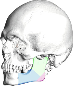
To better understand the jaw, it needs to be divided into the mandibular body, the mandibular angle and the ascending ramus ( Fig. 8.3 ). Generally speaking, the male jaw is larger, with greater body height and a broader ascending ramus. The mandibular angle formed by the body and ramus tends to be more acute in males, at times with everted gonial angles. The greater bone volume and vertical height are important factors when planning mandibular reshaping techniques in FGCS.
The male chin tends to be more square-shaped, with more pronounced and defined transitions between the chin and mandibular body, greater bone volume, and a more significant vertical dimension. Gender does not necessarily determine the position of the chin; that is, it is possible to find retro-positioned or over-projected chins in both men and women. However, a well-defined and projected chin may improve the overall aesthetics of the jaw-chin region.
Thyroid Cartilage (Adam’s Apple)
The larynx structure, which plays a key role in basic life processes like breathing and phonation, has a greater volume and is larger (greater in diameter and longer) in males. This structure should never be approached with the idea of feminizing it, since this would pose the unacceptable and unnecessary risk of damaging the vocal cords or even causing respiratory problems. Only the most prominent part of the thyroid cartilage should be modified. This allows for a significant reduction in the Adam’s apple without compromising its structural integrity.
Gender differences: secondary aspects
In addition to structural facial features, a series of secondary traits are equally important in the identification of facial gender. These include, most notably, the hair and hairline, facial hair, skin texture, and the distribution and volume of facial fat.
Male hair may be conditioned by androgenic alopecia (loss of hair due to hormonal influence) and tends to have an M -shaped primary hairline with recessions at the temples. The hairline of women usually has a rounded shape, their hair is not normally affected by alopecia, and, proportionally, the hairline implantation is higher in the center than in men. Although MtF transgender hairlines are comparable to male hairlines, they are distinctive in that alopecia stabilizes as a consequence of hormone treatment. The most common patterns found in MtF transgender patients are rounded (without recessions), M -shaped (receding hairline at the temples), and undefined (marked front line and temple recessions due to advanced alopecia). Hair density refers to the number of follicular units (FU) per square centimeter (FU/cm 2 ) on the scalp. Density and the composition of the FU can be easily measured with a simple handheld device called a densitometer. Additionally, the presence or absence of miniaturization can be assessed with a dermatoscope.
When approaching the upper facial third in MtF transgender patients, both the anatomy of the frontonasoorbital region and the overall condition of the hairline—format, height, and hair density—should be considered as a unit. For a better clinical analysis and broader understanding we have established an MtF hairline classification based on the observation and analysis of the hairlines of every transgender patient treated by our team through December 2015, a total of 492 patients. The analysis establishes five possible hairline height and format types: type I, hairline with normal height and rounded format; type II, hairline with normal height and receding hairline at the temples, often called an M -shaped hairline; type III, naturally high hairline; type IV, high hairline due to alopecia, which is usually associated with a receding hairline at the temples; and type V, undefined hairline due to advanced alopecia ( Table 8.1 ).
Almost all men have facial hair, which to a large extent conditions their skin type and quality, making it thicker and rougher. For many patients, facial hair is an important determining factor in their transition process.
The distribution and volume of facial fat is equally influenced by hormones. Women have a greater volume of facial fat with the distribution more concentrated in the middle third of the face (cheek area).
Since all of these features can be heavily determined by hormones, they generally respond well to hormone therapy. Conceptually, secondary features play an important role in determining facial gender and it is therefore preferable to treat them before beginning structural FGCS (at least 6 months before surgery) ( Fig. 8.4 ). Prior hormone therapy and FGCS is a combination that improves the results obtained with surgery.

Facial Feminization Procedures: Techniques, Goals, and Considerations
This section describes the main procedures that comprise FGCS. For a better understanding, the face is divided into four key areas and the most important procedures for each area are discussed ( Table 8.2 ).
The upper third: forehead and hairline
Despite the fact that the frontonasoorbital complex is one of the main areas that determines the identification of facial gender, the hairline also plays a crucial role in the upper third of the face. The combined evaluation of these two features should be a basic premise of FGCS.
Forehead Reconstruction
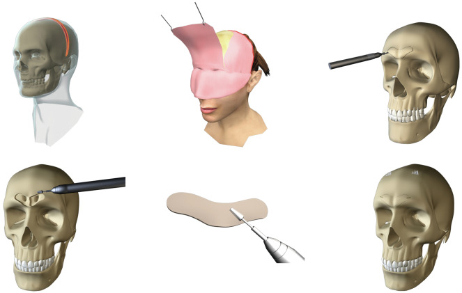
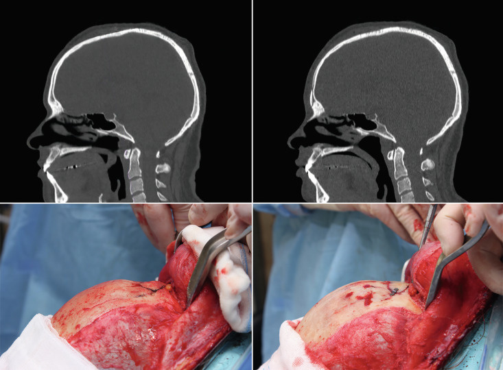
This is one of the basic procedures in facial feminization. It completely modifies the frontonasoorbital region and softens and feminizes the patient’s expression. The surgical plan is devised to open the frontonasal angle, retroposition the anterior wall of the frontal sinus, open the orbital areas, soften the entire forehead surface, and reposition the eyebrows above the new supraorbital ridge, while always maintaining the anatomical integrity of the entire area ( Figs. 8.5 and 8.6 ). The sequence in Fig. 8.7 provides a step-by-step description of the reconstruction technique proposed by our team. Despite the fact that other authors defend different techniques (isolated burring, the use of filling materials), in our experience, the proposed reconstruction technique offers satisfactory and safe results regardless of the anatomy of the frontal region ( Fig. 8.8 ).
Finally, it is important to discuss the best access (approach route) to reach the frontal bone region: modified coronal approach (anterior or posterior) or hairline approach. In our opinion, this access should be based on the characteristics of the patient’s hairline and its implantation (the distance from the nasal root to the beginning of the hairline).
Hairline Treatment
The hairline is a basic element in the identification of facial gender and, therefore, must be addressed to obtain a satisfactory and natural result in the upper third of the face. There are two options for treatment of the hairline. First, the hairline can be redefined using a hair transplant technique. The main areas to treat with a hair transplant are the receding hairline at the temples; however, the central section can also be addressed if the density is poor or if a small advancement (up to 1 cm) of the hairline is desired. This is recommended for patients with an M -shaped hairline, with sufficient hair density, and without active androgenetic alopecia (or with stabilized alopecia due to hormone treatment—type II MtF hairline). Depending on how the hair follicles are obtained, either follicular unit strip surgery (FUSS) or follicular unit extraction (FUE) can be used. In the FUSS technique the follicles are obtained from a strip of scalp removed in a surgical procedure, while in the FUE technique the follicles are obtained one by one, without any need for an invasive surgical process. This latter technique usually requires more experience given its technical complexity and it generally takes longer. The new hairline is designed to look natural, paying attention to parameters such as density and unevenness.
An alternative treatment to hair transplantation involves a hairline lowering surgery (HLS). The objective of HLS is twofold: to decrease the overall height of the forehead and to serve as an access point to reconstruct the frontonasoorbital complex (if required). This is only recommended for patients with a significantly and disproportionately high hairline (type III MtF hairline). A maximum of 2.5 cm of skin is removed and a 2 mm incision (future scar) is made above the hairline. Resorbable anchors (Endotine Forehead-mini device, Coapt Systems Inc., Palo Alto, CA, USA) can be placed to facilitate the advancement and eliminate the tension between the edges of the wound, helping to improve scarring. The lateral extension of the incision is hidden in the hair, since advancement in this area is not an objective of the surgery.
In most cases, there are a number of disadvantages to this technique: (1) the possibility of leaving a visible scar in a highly exposed part of the face; (2) the possibility of leaving an excessively short forehead in the center region, which could produce unnatural results; and (3) potentially limited results if surgical closure of the side temples is attempted due to excessive tension in the scarring area. In our experience, only 1 out of every 20 patients is a candidate for this type of approach and treatment. However, and despite the almost total lack of relevant bibliographic references, our professional experience suggests that this is the approach most commonly used around the world to treat the hairline in FGCS.
Forehead Reconstruction and Simultaneous Hair Transplant
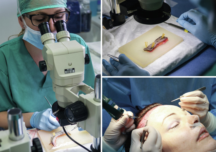
If the patient is a candidate for hairline treatment via hair transplant and also a candidate for forehead reconstruction, our team has developed a simultaneous hair transplant (SHT) technique. This technique consists of taking advantage of the strip of scalp obtained in the modified coronal approach, which we have used to access the frontal region. This allows us to harvest the hair follicles on this strip in the same way that they are obtained with the conventional FUSS transplant technique described above. Once the forehead reconstruction is done, a new hairline is designed and the hair follicles obtained are grafted in place (there are an average of 2000 FU per strip, meaning some 3900 hairs). To reduce the risks associated with prolonged general anesthesia, the patient is woken up and kept under light sedation for the duration of the SHT procedure. Thanks to this technique, the entire upper third can be treated as part of the same surgical process, which is highly advantageous for many patients ( Figs. 8.9 and 8.10 ). Androgenic alopecia must be completely stabilized before this technique can be used. In cases where there has been notable hair loss from the area where the strip of scalp would normally be obtained, we can simply position the coronal incision further back, even in the occipital region if necessary (posterior). The number of follicles that can be obtained from the strip is limited, so if the result of the SHT does not fully meet the objective of closing the temple recession, or if more density of hair is required, a second standard hair transplant procedure (FUSS or FUE) can be performed some months later (see Table 8.1 ).
The middle third: cheeks, nose, and upper lip
Malar Augmentation
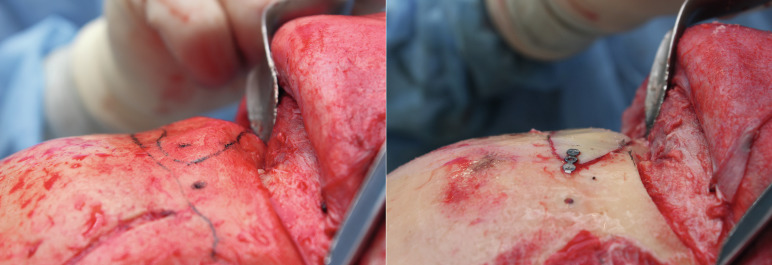
Although various cheek augmentation alternatives exist, we propose two options based on our experience. The first involves the use of rigid implants that are fixed to the bone using osteosynthesis material (positioning screws) to ensure stability ( Fig. 8.11 ). When necessary, they can be customized to the patient’s specific needs. They must be placed via an intraoral approach. The results are quite stable over time. If the volume of the implant is not carefully considered, the results may be artificial.
Related posts:
 Mental Health Care for the Adult Transgender Patient
Mental Health Care for the Child and Adolescent Transgender Patient
Hormone Treatment for the Adult Transgender Patient
Primary and Preventative Care for Transgender Patients
Gynecologic Care for Transgender Patients
Hysterectomy for the Transgender Man
Mental Health Care for the Adult Transgender Patient
Mental Health Care for the Child and Adolescent Transgender Patient
Hormone Treatment for the Adult Transgender Patient
Primary and Preventative Care for Transgender Patients
Gynecologic Care for Transgender Patients
Hysterectomy for the Transgender Man
Stay updated, free articles. Join our Telegram channel

Full access? Get Clinical Tree


