Facelift with SMAS Flaps

Chapter 9 Facelift with SMAS Flaps


The history of surgical modifications of the face is perhaps as old as surgery itself and the historic record is unclear as to when and where the first ‘facelift’ was performed. By the early 20th century surgeons had begun performing procedures to rejuvenate the face and techniques consisting of small excisions of facial skin had begun to evolve. These initial procedures were limited however, because they were performed using small discontinuous incisions, and because no skin undermining was performed.


Summary


Introduction


The history of surgical modifications of the face is perhaps as old as surgery itself and the historic record is unclear as to when and where the first ‘facelift’ was performed. By the early 20th century surgeons had begun performing procedures to rejuvenate the face and techniques consisting of small excisions of facial skin had begun to evolve. These initial procedures were limited however, because they were performed using small discontinuous incisions, and because no skin undermining was performed.


In 1920 Bettman described the continuous temporal-periauricular incision plan that became the prototype of that used in contemporary procedures, setting the stage for more comprehensive and balanced improvements, and in 1927 Bames recognized that undermining skin could further enhance outcomes. These developments comprised the foundation of the classical facelift procedures that were subsequently performed without major modification over the next four decades.


The modern era of facelift technique began in 1974 when Swedish surgeon Torg Skoog described dissection beneath the superficial fascia of the face and neck, a tissue layer subsequently described anatomically by Mitz and Peyronie that ultimately became known as the SMAS (superficial musculoaponeurotic system). Dissecting the SMAS allowed sagging deeper facial tissues to be repositioned to a more youthful position by an inelastic fascial layer, and tension to be diverted away from the skin. This resulted in softer more natural appearances, improved scars, and a significant improvement in the longevity of the procedure.


Numerous modifications have been made to Skoog’s procedure, and his idea has spawned alternative ways in which the SMAS is used. Nonetheless, the concept that the SMAS is the tissue layer most closely associated with age-associated facial tissue ptosis and the most logical means by which to correct it forms the foundation of most modern facelift techniques.


Facelift techniques


Some of the advantages and disadvantages of different facelift techniques are listed in Table 9.1.


Table 9.1 Advantages and disadvantages of different types of facelift techniques


Facelift Advantages Disadvantages and complications
Skin only Conceptual and technical simplicity Secondary facelift deformities caused by skin tension
SMAS plication No sub-SMAS dissection so technically less demanding and time consuming than procedures in which a flap is raised Wide skin flap undermining; contour irregularities if sutures are not placed carefully; potential injury to facial nerve branches and parotid gland or duct; little if any midface improvement
SMASectomy No sub SMAS dissection required; skin and SMAS can be shifted along different vectors so skin tension can be set low, and sideburn displacement and shifting of cervical wrinkles onto the face can be minimized Wide skin undermining; facial nerve branches at risk when SMAS excised and sutured; may be contour irregularities; minimal improvement in midface
Deep plane Single-layer dissection so flap elevation is easier and less time consuming than multilayer dissection and flap is thicker with a better blood supply Potential excessive sideburn displacement, and shifting of cervical rhytids to the face; unnatural appearances caused by extraordinary tension; midface improvement not always as expected; facial nerve branches and other structures at risk
Composite Similar to those for a deep plane lift, but also allows repositioning of orbicularis oculi and raising of lid-cheek junction Similar to those of deep plane technique, but also may be prolonged periorbital edema and denervation of orbicularis oculi
Lamellar SMAS dissection and bidirectional Skin and SMAS advanced bidirectionally in different amounts along separate vectors, and suspended under differential tension so avoiding skin tension, hairline displacement, and wrinkle shifts Technique dependent and time consuming; flaps more fragile; facial nerve branches at risk
Extended SMAS Improved effect in the mid face and infraorbital region and increased support of the lower eyelid More technique dependent and time consuming; flap is more fragile; facial nerve branches at risk
High SMAS Restoration of youthful upper cheek contour; filling of infraorbital region; increased support of lower eyelid; improved correction of nasolabial fold; readily combined with midface fat injections Similar to those techniques using a sub-SMAS dissection
Subperiosteal Dissection is familiar to most plastic surgeons and is largely deep to facial nerves; avoids many problems associated with skin-only procedures; improvement in infraorbital and upper midface areas in some cases Generally longer recovery and often hard to obtain optimal improvement along jawline; problems of suspension sutures
Endoscopic Minimal scarring No means for skin excision; little improvement in lower cheek and along jawline
Midface To improve midface area Steep learning curve and fraught with complications, especially when performed through a blepharoplasty incision, including lid retraction, ectropion, canthal displacement and dry eye; disappointing results
Suture suspension Seeming simplicity; easy marketability; can often be placed quickly under local or light anesthesia by inexperienced surgeons without surgical training; small incision; lower risk of hematoma, flap compromise and related complications Support of the face cannot be predictably obtained; infection; extrusion; traction dimples; visible bowstringing; nerve injuries; facial dyskinesias; chronic pain syndromes; abnormal appearances during animation; a tight or choking feeling when used in the neck; palpable knots and sutures beneath the skin and occasional erosion of overlying skin
MACS (minimal access cranial suspension) Seeming simplicity; no formal SMAS dissection; can be performed under local or light anesthesia; a shorter scar; shorter operating time. Technique dependent and contour irregularities can result; problems associated with the placement of a stiff and rigid suture in the superficial layers of the face; injury to the parotid gland, parotid duct, facial nerves, and other deep facial structures
Short scar Appeal to patients Limited access to deep layer structures; redundant skin cannot be shifted along proper vectors; prevent skin from being properly excised
Mini-lift Appeal to patients Minimal improvement


Skin only


Although the excision of skin formed the foundation of the facelift procedure for the first 70 years or so after its inception, this has ultimately been shown to be ineffective and conceptually flawed, and there are compelling reasons (Box 9.1) not to rely on skin resection only when surgically rejuvenating the face.


Despite their inherent drawbacks, well-known weaknesses, and tendency to produce secondary deformities, however, skin-only facelifts remain popular largely because of their conceptual and technical simplicity.


SMAS


Repositioning ptotic facial tissue using the SMAS is arguably the single most important advance in facelift technique since the inception of the procedure and various surgeons have devised different strategies to use it to improve the outcome of procedures. Each has certain advantages and disadvantages, but all share advantages over traditional skin-only procedures as follows:


Deep plane


A deep plane facelift consists of a sub-SMAS dissection first described by Skoog and later popularized by Lemon and Hamra in which the cheek skin and SMAS are raised together in one layer as a unified flap (Skoog flap). In the deep plane procedure the SMAS was used to reposition the lower cheek and jowl and the midface was then said to be elevated by ‘extraordinary tension’ on the preauricular portion of the flap.


Advantages of the deep plane technique include:


Potential disadvantages are:


Hamra has abandoned the deep plane technique in favor of the composite technique (see below), and the term deep plane has since come to be used generically by most surgeons for any procedure where there is some kind of modification of the SMAS is performed.


High SMAS


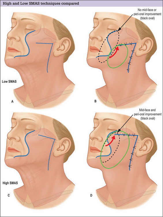
The conventional low cheek SMAS flap design in which the superior margin of the flap is planned below the zygomatic arch suffers the fundamental design flaw that it does not exert an effect on the tissues of the midface and infraorbital region. Low designs target the lower cheek and jowl only, and produce little if any improvement in the upper medial cheek area. Planning the flap higher, along the superior border of the zygomatic arch and making an extended dissection medially to mobilize midface tissue overcomes this problem and produces an improved result (Fig. 9.1A-D).


The high SMAS procedure can be performed using a two-layer lamellar technique with separate skin and SMAS flaps as advocated by Connell, or using a one-layer composite flap as practiced by Barton.


Advantages of a high SMAS plan include:


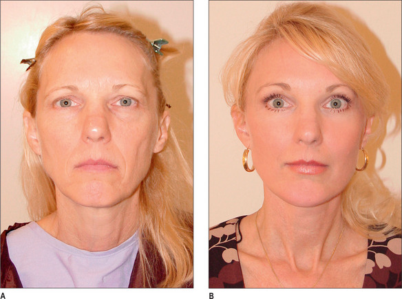
For many patients, the repositioning of midface tissue obtained with a high SMAS flap will be satisfactory in achieving the desired effect, and no additional or separate midface lift procedure will be required (Fig. 9.2A&B).


Disadvantages of the high SMAS technique are similar to other techniques in which a subSMAS dissection is made although some surgeons have expressed concern that the frontal branch of the facial nerve is at risk when high SMAS procedures are performed, they are chrucally safe and anatomically sound when performed as described.


Subperiosteal


Despite its seeming unsuitability, the periosteum has been advocated by Ramirez and others as the layer beneath which facelift dissection should be performed and the layer by which sagging superficial and deep facial tissues should be elevated.


Potential advantages of a subperiosteal facelift include:


The periosteum is, however, an intuitively and conceptually illogical choice for overall rejuvenation of the face because it is densely adherent to the facial skeleton and does not significantly descend as part of the aging process. It is also not closely associated with the intermediate layers of the face where most of the aging is known to occur.


Potential disadvantages of the subperiosteal technique include the following:


Endoscopic


Despite the importance of concealing scars in aesthetic surgery, other surgical specialties were the first to introduce and employ endoscopic techniques. The introduction and acceptance of the endoscopic fore-head lift eventually set into motion efforts to design a facelift performed using the same technique. Despite considerable effort, however, no proven or generally accepted technique for the face has emerged. In addition, focus has been diverted away from development of an endoscopic procedure to some extent by the introduction of techniques using percutaneously placed barbed threads and suspension sutures.


The major obstacle to the development and wide application of an endoscopic facelift is that skin redundancy remains a significant component of the aging deformity and although some support of deep layer tissue can be obtained, the endoscopic technique does not provide a means for this to be excised. Alternatives to excision including laser resurfacing are inadequate for all but a younger group of patients with small skin excess, and even these procedures have proven to be skin smoothing and not skin shrinking technologies.


For the most part the improvements obtained in endoscopic facelifts are limited largely to midface elevation, and improvements in appearance obtained from concomitantly performed forehead and neck surgery or the result of other ancillary procedures. Less improvement is obtained in the lower cheek and along the jawline.


Most endoscopic facelifts that have been proposed have been performed in a subperiosteal plane but often also attempt to resuspend the buccal fat pad, malar fat and the SOOF (sub-orbicularis oculi fat).


Midface


The midface is generally defined as an inverted triangular area situated over the anterior upper cheek that is bounded by:


In healthy, youthful appearing men and women this area is full and makes a smooth transition to the adjacent cheek and lower eyelid. As one ages however, there is generally atrophy of, and loss of volume from this area, and over time this results in an ill, haggard, and elderly appearance.


Midface atrophy is considered by some surgeons to be accompanied by descent of midface tissues, and the aging change occurring in the upper cheek area is as a result often, perhaps erroneously, referred to as midface ptosis.


The recognition of midface ptosis as a significant component of the changes occurring in the aging face, combined with the realization that the traditional SMAS facelift produced little or no improvement in the midface region, has led to a variety of procedures designed to specifically target the midface area. Many of these techniques are now being performed as isolated procedures or in conjunction with lower eyelid surgery.


Although there is merit in the idea of rejuvenating the midface, isolated midface lift procedures are still evolving, and have not been perfected. Most procedures have a steep learning curve and have been fraught with complications, especially when performed through a blepharoplasty incision. These complications include lid retraction, ectropion, canthal displacement and dry eye problems. As a result, many midface lift techniques have come to incorporate potentially problematic aggressive adjunctive surgical maneuvers including can thotomies, canthoplasties and orbicularis oculi muscle suspensions to prevent these problems. These maneuvers often result in a changed look that is disturbing to many patients, however, and carry a high risk of significant and troublesome complications of their own.


Many other valid arguments can be made against the use of isolated midface procedures including the fact that attractive rejuvenation of the face is possible without complete effacement of the nasolabial fold. Most patients with nasolabial folds had them when they were young and their presence reflects the individual’s anatomic make-up, and not an age related undesirable change. In such cases complete effacement is neither necessary nor desirable because they are an inherent and appropriate feature of the patient’s face.


As a practical matter, most of the improvement obtained by midface lifts consists only of elevation of the lid-cheek junction, and not correction of the nasolabial fold. Midface lifts also typically produce a subtle improvement that is difficult to document in photographs and not always noticed or appreciated by the patient. Because of this, it can be argued that time in the operating room is better spent on lower risk maneuvers that result in more noticeable improvement of higher priority to patients.


Careful examination of most patients who need or request a midface lift will show that they also need a facelift, and it is rare to encounter a patient with midface aging who does not also have sagging in the cheek and jowl. Because of this midface lifts are arguably more logically performed in conjunction with a formal facelift procedure. When this is done improvement is more balanced and comprehensive, and a more harmonious and natural appearing result is usually obtained. Healing is also generally faster and complications are less likely if midface improvement can be obtained through the facelift incision, rather than through a blepharoplasty or intraoral approach.


Most midface lifts are also generally conceptually flawed in that they mistakenly assume the problem seen in the anterior upper cheek is solely one of tissue sagging. Failure to acknowledge that significant atrophy is usually present has led to general disappointment following many procedures for both patients and surgeons, and has resulted in the addition of dermis fat grafts, orbital fat transposition and septal resets to midface lift procedures. It is questionable and remains to be answered however, whether these procedures can produce a restoration of lost volume as simply, naturally and effectively as obtained with fat injections.


Suture suspension


A variety of suture-lift procedures have been devised in which attempts are made to suspend deep facial tissues with sutures, including those in which:


Advantages of suture-lift procedures include:


Disadvantages of suture-lift procedures include:


Research and development continue on suture lift techniques, and it remains to be seen whether current problems can be solved. The fundamental problem in these procedures is that one must rely on a small rigid thread to support a relatively large area of delicate, inherently soft, mobile facial tissue. Over a short period of time these sutures typically cheese-wire through tissues they were intended to support, and can injure important neuromuscular structures. Suspension sutures also prevent natural depression and downward movement of facial tissues, impeding certain expressions and creating unnatural sensations and appearances.


The second major obstacle to be overcome is that although some support of deep layer tissue might be obtained, skin redundancy remains a significant component of the aging deformity for many patients, and suspension techniques do not provide a means for this to be excised. Alternatives to excision including laser resurfacing are inadequate for all but a younger group of patients with small skin excess, and even these procedures have proven to be skin-smoothing and not skin-shrinking technologies. As with endoscopic facelifts, a breakthrough in biomedicine may change this.


MACS lift


The MACS (minimal access cranial suspension) technique was developed and popularized by Tonnard and Verpaele for patients in the European market who were thought to be reluctant to undergo longer and ostensibly more complicated ‘traditional’ facelift procedures that were being performed in the Americas and elsewhere.


The MACS lift combines features of short-scar procedures with the concept of suture suspension of deep facial tissue. Tonnard and Verpaele assert the procedure shares little with traditional versions of these procedures, and clinical evidence indicates that when their technique is used outcomes are improved.


The MACS technique overcomes traditional limitations of suture suspension procedures in two important ways.


The MACS procedure has evolved and has been refined from its original description in which one or two plication sutures were placed and non-absorbable permanent monofilament sutures were used. Multiple sutures are now generally placed to more specifically target the midface, cheek, jowl and lateral neck, and this is said to distribute improvement more uniformly over the face and produce a more natural and long-lived improvement. Softer, absorbable sutures have also been substituted for the stiffer more rigid non-absorbable sutures originally recommended.


Although the MACS lift is considered by many who perform it to be a short-scar procedure, the typical incision plan used shortens the scar in the occipital area, but results in an incision along the more exposed temporal hairline, which is more difficult to conceal. This is of questionable benefit to younger patients who are typically thought to be the best candidates for the procedure.


Potential advantages of the MACS technique include:


Potential disadvantages of the MACS technique include:


Mar 4, 2016 | Posted by in Aesthetic plastic surgery | Comments Off on Facelift with SMAS Flaps

Full access? Get Clinical Tree

Get Clinical Tree app for offline access