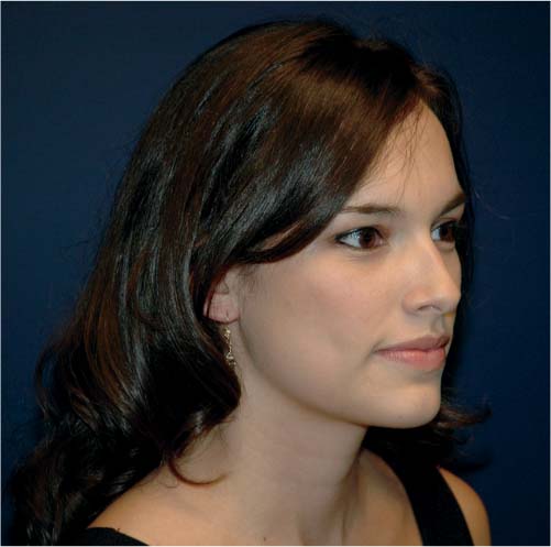
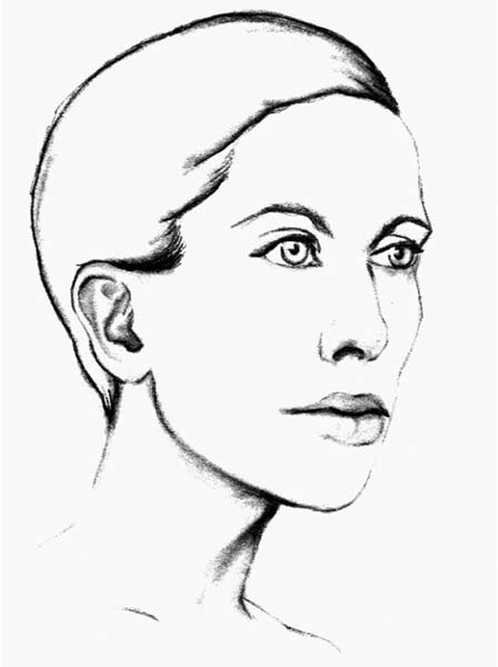
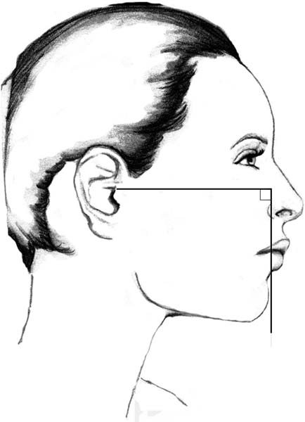
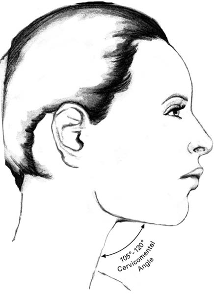
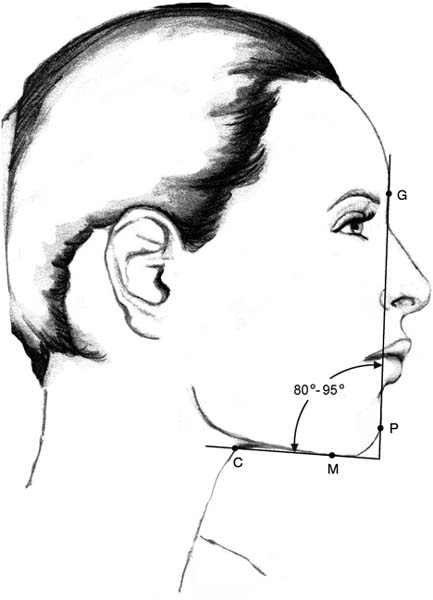
2 Accurately evaluating the face and neck can be one of the most challenging aspects of facial plastic surgery. The face can be analyzed in terms of aesthetic subunits, consisting of the forehead, the nose, and the eyes and brows. Ideal relationships of the facial subunits to one another can be described by using the nasofacial, nasofrontal, and other angles. Although techniques used to analyze the face have gotten considerable attention, the same cannot be said for evaluation of the neck. The relative paucity of articles dedicated to analyzing cervical deformities is surprising given the dramatic impact that an unaesthetic neck can have on facial appearance. The ability to successfully treat the neck depends largely on accurate preoperative analysis. A comprehensive cervical evaluation incorporates knowledge of anatomy, aesthetic ideals, and common deformities. This evaluation includes assessment of the skin, fat accumulation, soft tissue, and ligamentous and skeletal support of the neck. The initial consultation for plastic surgery of the neck has the three primary goals of: (1) assessment of deformity, (2) preoperative strategy, and (3) detailed communication, with the establishment of appropriate patient expectations. During this encounter the patient’s facial and cervical appearance are discussed and possible surgical solutions are outlined. Accurate, systematic analysis of the neck will guide the facial plastic surgeon to a correct surgical strategy for each patient. This chapter discusses methods used to assess the anatomy and aging-related changes of the neck. The youthful neck consists of firm, elastic soft tissue. The skin is taut and contours to the topography of the neck. The soft tissues of the youthful face are well-supported and do not sag or hang into the neck. A youthful, nonsurgically treated neck is shown in Figure 2.1. The mandibular border is well defined and the chin is well projected. The cervico-mental angle (CMA) is acute. The muscles and cartilages of the neck are defined by gentle contours. The neck is free of rhytids, platysmal banding, and jowl overhang. Comprehensive cervical evaluation can be simplified by assessing the appearance of discrete anatomical regions. According to Ellenbogen and Karlin, the aesthetic neck should be evaluated for a well-defined CMA, definition of the inferior mandibular border, a visible anterior border of the sternocleidomastoid (SCM) muscle, a subhyoid depression, and a thyroid bulge.1 Two important considerations not addressed by Ellenbogen and Karlin are mental projection and position of the hyoid bone. Fig. 2.1 A youthful neck untreated with surgery. The soft tissues of the face are elevated and well supported. The inferior mandibular border is defined as an uninterrupted shadow from the menton to the angle of the mandible. The CMA is acute and without significant accumulation of fat. The neck is free of rhytids, platysmal banding, and prominent submandibular glands. The mentum should be evaluated for anterior, lateral, and inferior projection. This should reveal a sculpted, well-de-fined border on both anterior and profile views.2 A projected mentum combined with an acute CMA visually separates the face from the neck. Photographers and artists accentuate this separation by casting a shadow into the submental triangle and anterior neck (Fig. 2.2). A weak, receding chin creates the illusion of a short neck. In a series of chin augmentations, Courtiss demonstrated that augmenting a deficient mentum can improve the aesthetic balance of the neck.3 Anteroposterior mental deficiencies (horizontal microgenia) are easily augmented with alloplasts. Chin implants are significantly less effective in correcting vertical and transverse deformities. For more complex deformities of the chin, bony osteotomy of the chin (genioplasty) is indicated. Evaluation of the chin should focus on its appearance in three-dimensional view. When viewed in profile, the ideal projection of the chin can be estimated by dropping a vertical line from the vermilion border of the lower lip (Fig. 2.3).4 In the aesthetic neck, the pogonion reaches this vertical line. The mental soft tissue will fall short of this line with microgenia or retrognathia. The ideal projection of the chin is gender-specific. It is acceptable for the pogonion in men to extend slightly beyond the ideal vertical line and for the pogonion in women to be slightly positioned behind this line. The more superior and posterior the hyoid bone, the more acute and more attractive the CMA. An inferiorly, anteriorly positioned hyoid bone can create an obtuse cervicomental deformity in the absence of microgenia, accumulation of submental fat, or platysmal laxity. Malpositioned hyoid bones are commonly referred to as “problem necks.” Intra-operative repositioning of the hyoid bone is challenging and typically requires transecting the submental musculature. Without myotomy, the patient should be counseled about the anatomical limitations of surgery to reposition the hyoid bone. Fig. 2.2 Accentuated submental shadow used to visually separate the face from the neck. Fig. 2.3 Ideal chin projection can be estimated by dropping a vertical line from the vermilion border of the lower lip. In the aesthetic neck, the pogonion reaches this vertical line. It is acceptable for the pogonion to extend slightly beyond this line in men and to fall slightly short of it in women. Guyuron reviewed dozens of cephaloxerograms and related hyoid-bone position to attractive and unflattering CMAs.5 He concluded that in a balanced neck the caudal border of the hyoid bone is at or above the level of the menton (the most inferiorly projecting point of the chin). An anteriorly, inferiorly located hyoid bone will limit the ability to surgically create a well-contoured submental triangle. The CMA is created by connecting a horizontal line extending through the menton with an oblique line following the anterior border of the neck (Fig. 2.4). The ideal CMA is between 105 and 120 degrees. When present in aesthetic proportions, this angle reveals a harmonious separation of the face and neck. The lower one-third of the face appears projected and the neck elongated. Powell and Humphrey described this anatomical region in terms of a mentocervical angle (MCA) (Fig. 2.5).6 The MCA is formed by the intersection of a vertical line drawn from the glabella (G) (the most prominent point of the forehead in a profile view) to the pogonion (P) (the anteriormost point of the chin) with a horizontal line connecting the menton (M) to the cervical point (C) (the innermost point between the submental area and the neck). The ideal MCA is between 80 and 95 degrees. An obtuse MCA blurs the distinction between the face and neck. The CMA and MCA are two of many means used to evaluate the submental triangle. Dedo chose to avoid mathematical analysis and described the ideal submental triangle as flat.7 A submental convexity will create the illusion of a short neck. A submental concavity will skeletonize the musculature in the floor of mouth. Submental accumulation of fat, laxity of the platysma muscle, an anteriorly located hyoid bone, and microgenia are all conditions affecting the submental triangle. It is important that the surgeon specifically identify the condition (s) contributing to an unattractive neck. Only then will the surgeon be able to accurately treat the responsible deformity. Fig. 2.4 The CMA is the angle created between the chin and the neck. The ideal CMA is between 105 and 120 degrees. Fig. 2.5 The MCA is formed by the intersection of a vertical line drawn from the glabella to the pogonion with a horizontal line connecting the mentum to the cervical point. The ideal MCA is between 80 and 95 degrees. The top of the neck is formed by a well-defined mandibular body and a prominent chin. A mandible with ideal height, length, and projection will create an angular jaw–neck contour. The inferior border of the mandible is lined by a shadow from the bony mentum to the mandibular gonion. This line should be free of blunting from ptosis of the submandibular gland and uninterrupted by jowl overhang. To improve the definition of the mandibular border, liposuction can be used above and below the mandible; however, a strip of subcutaneous fat should be left along the mandibular border to highlight the border itself.8 The ideal bony mandibular angle is well-defined and projected both laterally and inferiorly. Gonial deficiency can be corrected with a bone implant that wraps around the posterior and inferior borders of the mandibular angle. A sculpted posterior border of the angle distinguishes the face from the SCM muscle.
Evaluation of the Anatomy and Aging-Related Changes of the Neck
Criteria for the Aesthetic Neck
Point One: Mental Prominence
Point Two: Hyoid Position
Point Three: Cervicomental Angle
Point Four: Definition of the Mandibular Border
Stay updated, free articles. Join our Telegram channel

Full access? Get Clinical Tree


