Elevated/Brown-Black/Multicolored
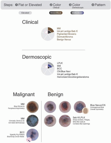 FIGURE 15.1 Color wheel: elevated/brown-black/multicolored. |
Step 1: Is the lesion flat or raised? Elevated
Step 2: What color is the lesion on clinical assessment? Brown/Black
Step 3: What is the dermoscopic color? Multicolored
Step 4: Is further elucidation needed to decide whether to biopsy or not? Yes
Is this a malignant or benign pattern?
Take a look at the color wheel in Figure 15.1.
When we elevate the clinically brown/black, dermoscopically multicolored lesions, we don’t change the differential from the flat lesions. However, we do now include a more advanced stage of malignant melanoma. These will be clinically obvious lesions for which dermoscopy is not needed.
Our benign lesions include LPLK and seborrheic keratosis and congenital/blue nevi.
Our malignancies include malignant melanoma again, as well as clinically obvious nodular melanoma. We also will see pigmented basal cell again.
Benign Lesions
Congenital/Combined Nevi
Pearls
Elevated/Brown-Black/Multicolored
These will have been present since birth.
May wobble with contact.
Step 4 Pattern: Remember to look for your Chapter 1 Patterns
Any of the melanocytic patterns: homogenous, globular/cobblestone, or reticular.
Shades of brown are not considered multicolored!
Sometimes, you may see a few visible comma vessels and milia-like cysts.
Bottom line: Benign, biopsy not necessary.
Examples
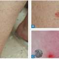
Figures 15.2 and 15.3 show a clinically elevated, brown/black lesions (A, B), with a dermoscopically multicolored (brown + other = gray, pink, yellow) pattern (C). These lesions are difficult to appreciate, but overall, we see a homogenous pattern with black, brown, and some blue. This lesion wobbles with contact, unless it has undergone a lot of rubbing over the years and has fibrosed to some extent. A clinical history indicates that the lesion has been present since birth and it resembles nearby lesions. Therefore, this lesion is unlikely to be malignant. Additionally, nodular melanomas are very fast growing, which would not fit this clinical picture. Diagnosis: Congenital/combined nevi.
Bottom line: Benign, biopsy unnecessary.
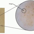
Figure 15.4 shows a clinically elevated, brown/black lesion (Figure 15.4A, B), with a dermoscopically multicolored (brown + other = gray, pink, yellow) pattern (Figure 15.4C). This lesion is an example of a congenital nevus. You can appreciate the dermoscopic dot/globular pattern, as well as some darker dots on the top of the network. Diagnosis: Congenital nevus.
Bottom line: benign, biopsy unnecessary.
Figure 15.5 shows a clinically elevated, brown/black lesion (Figure 15.5A, B) with a dermoscopically multicolored (brown + other = gray, pink, yellow) pattern (Figure 15.5C). This lesion is an example of a congenital nevus. You can appreciate the dermoscopic dot/globular
and cobblestone pattern, which is specific to congenital lesions. This lesion will wobble when in contact with the scope. Diagnosis: Congenital nevus.
and cobblestone pattern, which is specific to congenital lesions. This lesion will wobble when in contact with the scope. Diagnosis: Congenital nevus.
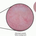
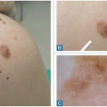
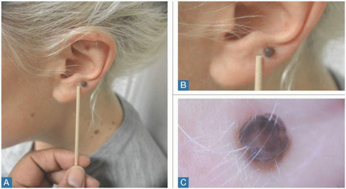 FIGURE 15.2 These clinically elevated lesions are dark brown, with a multicolored (brown + other = gray, pink, and/or yellow) dermoscopic pattern. Review these clinical and dermoscopic examples of a congenital nevus. A,B: Clinical examples. C: The dermoscopic example shows that a pattern is difficult to appreciate, but overall, it is homogeneous with black, brown, and even blue. This lesion should wobble when a contact scope is applied to it, but when lesions have been rubbed and fibrosed, it may be difficult to appreciate this feature. A clinical history will indicate that the lesion has been present since birth, resembles other lesions on the patient, and is clinically well circumscribed. The reality that nodular melanomas are very rapidly growing lesions also helps with the diagnosis of dark and multicolored congenital nevi. |
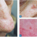
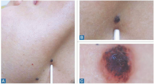 FIGURE 15.3 These clinically elevated lesions are dark brown, with a multicolored (brown + other = gray, pink, and/or yellow) dermoscopic pattern. Review these clinical and dermoscopic examples of a congenital nevus. A,B: Clinical examples. C: The dermoscopic example shows a pattern that is difficult to appreciate, but overall it is homogeneous with black, brown, and even blue. This lesion should wobble when a contact scope is applied to it, but when lesions have been rubbed and fibrosed, it may be difficult to appreciate this feature. A clinical history will indicate that the lesion has been present since birth, resembles other lesions on the patient, and is clinically well circumscribed. The reality that nodular melanomas are very rapidly growing lesions also helps with the diagnosis of dark and multicolored congenital nevi. |
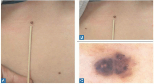 FIGURE 15.4 These clinically elevated lesions are dark brown, with a multicolored (brown + other = gray, pink, and/or yellow) dermoscopic pattern. Review these clinical and dermoscopic examples of a congenital nevus. A,B: Clinical examples. C: The dermoscopic example shows a dot/globular pattern. You can also see dark dots on the network. |
Bottom line: Benign, biopsy unnecessary.
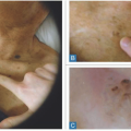
Figure 15.6 shows a clinically elevated, brown/black lesion (Figure 15.6A, B) with a dermoscopically multicolored (brown + other = gray, pink, yellow) pattern (Figure 15.6C). This lesion is an example of a congenital nevus. Again, we can see the dermoscopic dot/globular pattern.
The erosion or bleeding seen in this lesion is most likely due to outside trauma. Additionally, this lesion wobbles when in contact with the scope. Diagnosis: Congenital nevus.
The erosion or bleeding seen in this lesion is most likely due to outside trauma. Additionally, this lesion wobbles when in contact with the scope. Diagnosis: Congenital nevus.
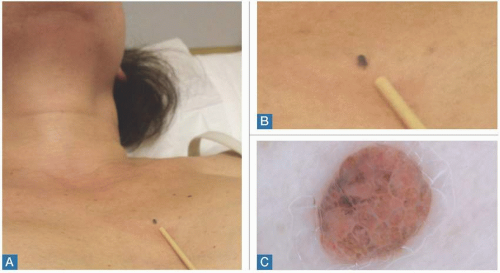 FIGURE 15.5 These clinically elevated lesions are dark brown, with a multicolored (brown + other = gray, pink, and/or yellow) dermoscopic pattern. Review these clinical and dermoscopic examples of a congenital nevus. A,B: Clinical examples. C: The dermoscopic example shows a dot/globular and cobblestone pattern that is very specific to congenital lesions. It is more difficult to see the comma-like vessels in darker lesions. Additionally, when a contact scope is applied to the lesion, it wobbles. |
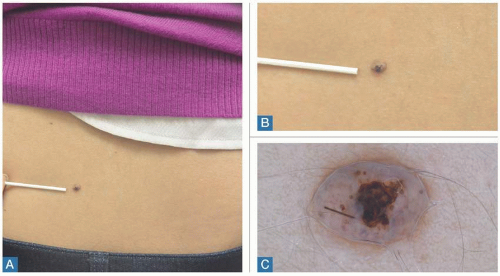 FIGURE 15.6 These clinically elevated lesions are dark brown, with a multicolored (brown + other = gray, pink, and/or yellow) dermoscopic pattern. Review these clinical and dermoscopic examples of a congenital nevus. A,B: Clinical examples. C: The dermoscopic example shows a dot/globular pattern. Note the erosion/bleeding seen in this lesion, most likely due to outside trauma. Additionally, when a contact scope is applied to the lesion, it wobbles. |
Bottom line: Benign, biopsy unnecessary.
Figure 15.7 shows a clinically elevated, brown/black lesion (Figure 15.7A, B), with a dermoscopically multicolored (brown + other = gray, pink, yellow) pattern (Figure 15.7C). This
is an example of an elevated blue nevus. Here, we see the homogenous blue symmetric pattern that begins to have a blue-white veil appearance because of its elevation. This makes it more difficult to differentiate from a nodular melanoma. The clinical history, and lack of other malignant features often seen with the more rapidly growing and aggressive nodular melanomas, helps to differentiate these lesions. New blue nevi that develop on the scalp, such as this one, are often biopsied due to reported cases of these lesions becoming locally aggressive. Diagnosis: Elevated blue nevus.
is an example of an elevated blue nevus. Here, we see the homogenous blue symmetric pattern that begins to have a blue-white veil appearance because of its elevation. This makes it more difficult to differentiate from a nodular melanoma. The clinical history, and lack of other malignant features often seen with the more rapidly growing and aggressive nodular melanomas, helps to differentiate these lesions. New blue nevi that develop on the scalp, such as this one, are often biopsied due to reported cases of these lesions becoming locally aggressive. Diagnosis: Elevated blue nevus.
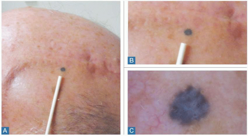 FIGURE 15.7 These clinically elevated lesions are dark brown, with a multicolored (brown + other = gray, pink, and/or yellow) dermoscopic pattern. Review these clinical and dermoscopic examples of a blue nevus. A,B: Clinical examples. C: The dermoscopic example shows a symmetric homogeneous blue that begins to have a blue-white veil appearance when elevated. This appearance makes it more difficult to distinguish from nodular melanoma. The clinical history, and lack of other malignant features often seen with the more rapidly growing and aggressive nodular melanomas, helps differentiate these lesions. New blue nevi of the scalp are often biopsied, due to reported cases of these lesions becoming locally aggressive.
Related posts:Stay updated, free articles. Join our Telegram channel
Full access? Get Clinical Tree
 Get Clinical Tree app for offline access

|