Elevated/Brown-Black/Brown
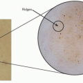
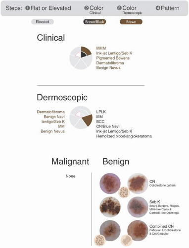 FIGURE 13.1 Color wheel: elevated/brown-black/brown. |
Step 1: Is the lesion flat or raised? Elevated
Step 2: What color is the lesion on clinical assessment? Brown-Black
Step 3: What is the dermoscopic color? Brown
Step 4: Is further elucidation needed to decide whether to biopsy or not? Sometimes
Take a look at the color wheel in Figure 13.1.
What’s missing on the differential? Malignant lesions!
If you evaluate a lesion and determine that it is a clinically elevated, brown-black, and dermoscopically brown lesion, you are dealing with a benign lesion. You will not need to biopsy these lesions.
Our differential includes only benign entities, congenital and combined nevi, as well as seborrheic keratosis.
There are no malignancies on our list. By definition, malignancies that are elevated have already progressed to an advanced stage and will be clinically suspicious. If any of these elevated lesions have more atypical features, and truly do not resemble their neighbors, a biopsy should be done. However, this category would be an exception to the rule.
Congenital or Combined Nevi
Pearls
Elevated/Brown-Black/Brown
Will have been present since birth
May wobble with contact
Step 4 Pattern: Remember to review your Chapter 1, Patterns.
You can see all of the benign melanocytic patterns: homogenous, globular/cobblestone, or reticular.
Shades of brown should not be considered multicolored! The darkest shade seen in the lesion will classify which color wheel category to choose.
Sometimes you can see a few visible comma vessels and milia-like cysts.
Bottom line: Benign, biopsy not necessary.
Examples
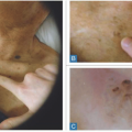
Figure 13.2 shows a clinically elevated, brown-black lesion (Figure 13.2A, B), with a dermoscopically brown pattern (Figure 13.2C). This is a classic example of a congenital nevus with a symmetric, diffuse globular pattern. Diagnosis: Congenital nevus.
Bottom line: Benign, biopsy unnecessary.
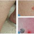
Figure 13.3 shows a clinically elevated, brown-black lesion (Figure 13.3A, B), with a dermoscopically brown pattern (Figure 13.3C). This is a classic example of a congenital nevus with a symmetric, diffuse globular pattern. This lesion will wobble with contact. Diagnosis: Congenital nevus.
Bottom line: Benign, biopsy unnecessary.
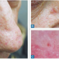
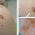
Figure 13.4 shows a clinically elevated, brown lesion (Figure 13.4A, B), with a dermoscopically brown pattern (Figure 13.4C). This is a classic example of a compound nevus. You can appreciate comma-like vessels and faint pigmentation. The lesion also wobbles. Often, there is an associated reticular network with compound lesions, but you cannot see this more superficial pattern in this example. Diagnosis: Compound nevus.
Bottom line: Benign, biopsy unnecessary.
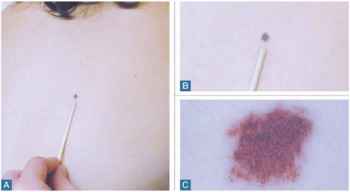 FIGURE 13.2 These clinically elevated lesions are dark brown, with a brown dermoscopic pattern. Review these clinical and dermoscopic examples of a melanocytic nevus. A,B: Clinical examples. C: The dermoscopic example shows a dot/globular pattern. |
Figure 13.5 shows an example of a two-toned lesion; it is best to choose the darkest clinical color for the color wheel. We can see a clinically elevated, brown-black lesion (Figure 13.5A, B) with a dermoscopically brown pattern (Figure 13.5C). This is a classic example of a congenital nevus. You can appreciate a globular dot pattern. There is also an eccentric peripheral reticular pigmentation, but the lack of other malignant features in light of this lesion being clinically elevated and dark brown makes malignancy unlikely. Often, there is an associated reticular
network with compound lesions, but you cannot see this more superficial pattern in this example. Diagnosis: Congenital nevus.
network with compound lesions, but you cannot see this more superficial pattern in this example. Diagnosis: Congenital nevus.
Related posts:
Stay updated, free articles. Join our Telegram channel

Full access? Get Clinical Tree


