Identify the anatomy of the ear (Fig. 77-1).
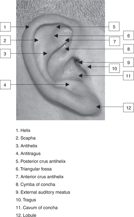
Figure 77-1 Anatomy of the ear.
 Which embryologic structures give rise to the ear?
The first two branchial arches give rise to six hillocks that form the ear.
The first arch forms the malleus, incus, and anterior hillocks (1–3).
The second arch forms the stapes and posterior hillocks (4–6).
 What structures arise from the anterior (1–3) hillocks?
The tragus, root helix, and superior helix.
 What structures arise from the posterior (4–6) hillocks?
The antihelix, antitragus, and lobule.
 What ear structures are formed from the first branchial groove?
The external auditory canal and concha.
 What is the average length of the ear?
The average length is 6 cm. Approximately 90% of the growth occurs by 5 years of age.
 What is the angle of protrusion of the ear?
25 to 30 degrees.
 What is the vascular supply of the ear?
The ear is supplied by branches of the external carotid artery:
1. The lateral ear is supplied by the superficial temporal and posterior auricular arteries.
2. The medial ear is supplied by the posterior auricular and posterior occipital arteries.
3. The triangular fossa and scapha are supplied by the helical branch of the superficial temporal artery.
4. The concha is supplied by septal perforators of the posterior auricular artery.
 What is the vascular supply to the ear cartilage?
The cartilage is an avascular structure, which is supplied by the surrounding perichondrium and soft tissue.
 What is the lymphatic drainage of the ear?
The lymphatic drainage may vary, although it generally follows embryologic development:
1. The superolateral ear and anterior external auditory meatus drain to the parotid nodes.
2. The superomedial ear and posterior external auditory meatus drain to the mastoid nodes.
3. The inferior ear and lower external auditory meatus drain to the superficial cervical nodes.
4. The concha and meatus drain to the preauricular nodes.
 Describe the innervation of the ear.
1. The auriculotemporal branch (V2) supplies the superolateral ear.
2. The great auricular branch (C2–3) supplies the inferolateral and inferomedial ear.
3. The lesser occipital branch (C2–3) supplies the superomedial ear.
4. The auditory (“Arnold’s”) branch (X) supplies the concha and external auditory canal.
 How do you anesthetize the ear?
Anesthesia of the ear, except the concha and external auditory canal, can be performed with a ring block around the base of the ear. Anesthesia to the concha and external auditory canal must be performed with local infiltration.
 What is the treatment for ear burns?
The primary goal is to prevent chondritis. Topical mafenide is used for dressing changes. The eschar is left in place as a biologic dressing. Defects with intact perichondrium will heal in, while others will require late excision and repair.
 What is the treatment for frostbite of the ear?
Topical mafenide is used for burns and frostbite. It is highly soluble, which allows penetration of eschar and cartilage. Sulfonomides are carbonic anhydrase inhibitors, which can lead to metabolic acidosis.
 What is the management of an ear hematoma?
Ear hematomas are treated by aspiration or incision and drainage. Bolster dressing is applied to prevent reaccumulation. Hematoma forms between the cartilage and perichondrium of the ear. This disrupts the vascular supply to the cartilage, which may become necrotic or infected. Clotted blood forms a fibrotic mass that deforms the overlying tissue. This is known as a “cauliflower,” “boxer’s,” or “wrestler’s” ear.
 What are the most common malignancies of the ear?
1. Squamous cell cancer: 50% to 60%
2. Basal cell cancer: 30% to 40%
3. Melanoma: 1% to 2%
 What are the causes of a prominent ear?
1. Underdeveloped antihelical fold, leading to protrusion of the scapha and helical rim, causing prominence in the upper to middle 1/3.
2. Prominent concha, causing middle 1/3 prominence.
3. Protruding earlobe, causing lower 1/3 prominence.
 What is a constricted ear?
Related posts:
Stay updated, free articles. Join our Telegram channel

Full access? Get Clinical Tree


