Dorsalis Pedis Free Flap for Eye Socket Reconstruction
B. GUYURON
H. P. LABANDTER
Contracted or missing eye sockets, especially in an irradiated orbit, represent a major reconstructive task for the orbital surgeon. Failure of simple eye socket reconstruction requires complex procedures. Most grafts are doomed to failure, and local flaps may leave undesirable donor-site scars. Distant flaps require many stages and leave visible scars, and the patient is placed in an uncomfortable position for 2 or 3 weeks. In selected patients, we have transferred the dorsalis pedis flap to reconstruct the missing or contracted eye socket.
INDICATIONS
This procedure should be used only if simpler techniques of eye socket reconstruction fail. The procedure of choice for eye socket reconstruction is the application of a full-thickness skin graft. If this fails, the next choice is a postauricular fasciocutaneous flap (see Chapter 30) or a secondary axial-pattern flap (1). If the clinical examination indicates a diminished or absent superficial temporal pulse and the local condition supports this finding, a vascularized dorsalis pedis flap can be chosen for eye socket reconstruction.
FLAP DESIGN AND DIMENSIONS
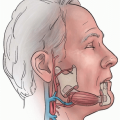
We use the central portion of the dorsalis pedis flap, the size of the flap depending on the size of the defect (Fig. 34.1A). Although the short and long saphenous veins can be used for the venous anastomosis, we prefer to use one of the venae comitantes of the anterior tibial artery because of its length and ease of dissection. The flap is designed with adequate dimensions to reconstruct the socket, as well as an additional de-epithelialized portion of the margin of this flap which is used to correct the periorbital deficit.
Related posts:
 Cheek Rotation Skin (Mustardé) Flap to The Lower Eyelid
 Wraparound Cartilage Flap for Correction of Cleft-Lip Nasal Deformity
 Oral Mucosal Flaps for Septal Reconstruction
 Postauricular and Retroauricular Scalping Flap (The Paras Flap)
 Platysma Musculocutaneous Flap to The Lower Lip
 Microvascular Free Transfer of Serratus Anterior and Rib Composite Flap
Stay updated, free articles. Join our Telegram channel

Full access? Get Clinical Tree


