, Shimin Chang2, Jian Lin3 and Dajiang Song1
(1)
Department of Orthopedic Surgery, Changzheng Hospital Second Military Medical University, Shanghai, China
(2)
Department of Orthopedic Surgery, Yangpu Hospital Tongji University School of Medicine, Shanghai, China
(3)
Department of Microsurgery, Xinhu Hospital Shanghai Jiao Tong University, Shanghai, China
Earley and Milner [1] described distally based first web flaps that included branches of the first dorsal and plantar metatarsal arteries and which they used to cover distal dorsal foot defects.
The reverse first dorsal metatarsal artery (FDMA) flap has a longer vascular pedicle and can be used for distal foot wounds [2].
30.1 Vascular Anatomy
The dorsalis pedis artery, which is an extension of the anterior tibial artery, supplies the flap. The anterior tibial artery lies lateral to the tibialis anterior tendon and medial to the extensor hallucis longus tendon at the entrance of the extensor retinaculum or the ankle. It courses under the retinaculum and emerges medial to the extensor hallucis longus tendon as the dorsalis pedis artery. The dorsalis pedis artery branches to form the arcuate artery and lateral and medial tarsal arteries, which supply structures beneath the extensor tendons and are not harvested as part of the flap [3–5].
The artery branches into the first dorsal metatarsal artery and deep perforating branch at the first intermetatarsal space. The critical point, where the first dorsal metatarsal artery (or the first plantar metatarsal artery in cases when the former branched from the latter) was located 10 mm distal to the tarsal-first metatarsal joint and 5.5 mm plantar from the dorsal surface of the second metatarsal bone (Fig. 30.1).
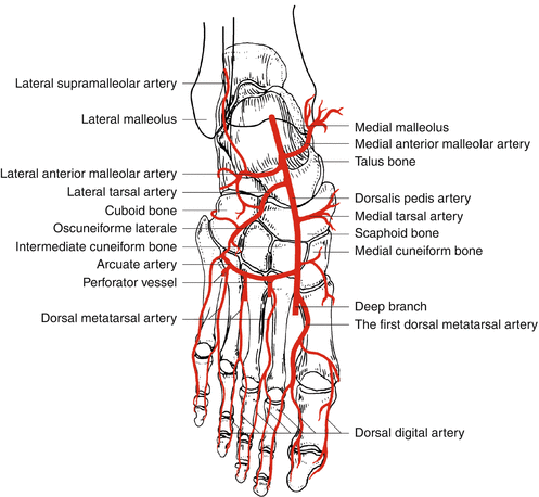
Fig. 30.1
Vascular anatomy of the dorsalis pedis artery
30.2 Illustrative Case
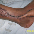
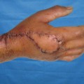
A 32-year-old man presented with a press injury over his distal portion of the foot and big toe, with skin and soft tissue necrosis. A thorough debridement was carried out, leaving a defect of 4.5 cm × 2.0 cm over the big toe (Fig. 30.2).
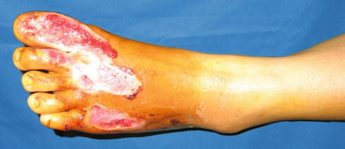
Fig. 30.2
Preoperative view
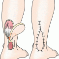
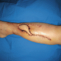
Flap Design
Related posts:

Stay updated, free articles. Join our Telegram channel

Full access? Get Clinical Tree


