48
Distal Interphalangeal Joint Dislocations
John D. Wyrick
History and Clinical Presentation
A 24-year-old right hand dominant man injured his right small finger while playing softball. The ball struck him on the tip of the finger and he thought he “only jammed” the finger. He describes pulling on the finger, which made it feel a little better, but always noted some deformity, which he attributed to swelling. He did not seek medical attention until 10 days after the injury. When seen, he described persistent pain and lack of motion as the main reasons for seeking help.
Physical Examination
Significant swelling diffusely throughout the right small finger is noted. It is more prominent distal to the proximal interphalangeal (PIP) joint. No wounds were present. Grossly, there was a prominence dorsally at the distal interphalangeal (DIP) joint and obvious deformity. Due to pain his passive motion was not tested, but his active motion was 10 to 25 degrees. Sensibility testing revealed static two-point discrimination of 5 mm.
Imaging Studies
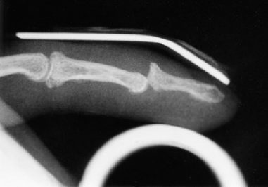
Biplanar radiographs showed a dorsal dislocation of the DIP joint. No fracture was present (Fig. 48–1).
Differential Diagnosis
DIP joint dislocation
Fracture of distal phalanx
Mallet injury
Avulsion of flexor digitorum profundus (FDP) tendon
PIP joint dislocation
PEARLS
- In a spontaneously reduced dislocation with an open wound, the joint must be explored and irrigated thoroughly.
PITFALLS
- Avoid chronic deformity and disability by testing active extension and flexion of the DIP joint after reduction.

Figure 48–1. Lateral radiograph after reduction attempt with persistent dorsal dislocation of distal interphalangeal (DIP) joint.
Diagnosis
Dislocation of the DIP Joint
Dislocations of the DIP joint are not commonly seen. More common is the dorsal PIP joint dislocation, which is the result of a hyperextension injury. Often these DIP joint injuries are reduced in the field and are never seen for medical attention.
Stay updated, free articles. Join our Telegram channel

Full access? Get Clinical Tree


