(1)
Department of Dermatology, Freiburg University Medical Center, Freiburg, Germany
8.1.1 Capillary Didymosis
8.1.1.2 Nevus Vascularis Mixtus, a Hallmark of Mixed Vascular Nevus Syndrome (Ruggieri-Leech Syndrome)
8.1.2 Cutis Tricolor
8.1.2.1 Ruggieri-Happle Syndrome
8.1.2.2 Cutis Tricolor Parvimaculata
Abstract
In the group of didymotic genodermatoses we can distinguish allelic from non-allelic twin spotting. Molecular proof is so far lacking but it is very likely that the concept will be confirmed in the proposed examples of allelic didymosis such as the paired occurrence of nevus flammeus and nevus anemicus, nevus vascularis mixtus representing the hallmark of a recently delineated neurocutaneous syndrome, various forms of cutis tricolor, and paired linear areas of either excessive or absent involvement as noted in Darier disease and epidermolytic ichthyosis of Brocq. By contrast, the possible cases of non-allelic didymosis should be considered with great caution since the etiological concept has now been disproved in phacomatosis pigmentokeratotica and phacomatosis cesioflammea. Future research may show whether the assumption of non-allelic didymosis will be confirmed or falsified in phacomatosis spilorosea or phacomatosis melanorosea.
The two dissimilar alleles causing didymotic skin disorders (see Sect. 3.1.1.5) may involve the same gene locus (allelic didymosis), or they may affect different loci (nonallelic didymosis) on either of a pair of homologous chromosomes.
8.1 Allelic Didymosis
The following disorders are suggestive of allelic twin spotting, but molecular proof is so far lacking.
8.1.1 Capillary Didymosis
This form of didymosis occurs rather frequently. From a clinical point of view, different types can be distinguished.
8.1.1.1 Nevus Flammeus Twinned with Nevus Anemicus
8.1.1.2 Nevus Vascularis Mixtus, a Hallmark of Mixed Vascular Nevus Syndrome (Ruggieri-Leech Syndrome)
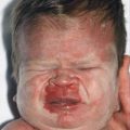
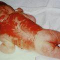
This phenotype represents an admixture of reticular telangiectatic lesions and angiospastic spots of nevus anemicus [7]. It occurs as an isolated skin disease (Fig. 8.2) or as a hallmark of a neurocutaneous syndrome characterized by distinct brain malformations (mixed vascular nevus syndrome, Ruggieri-Leech syndrome) [16, 22].
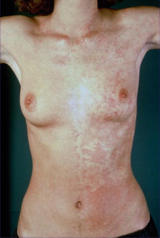
Fig. 8.2
Nevus vascularis mixtus (Courtesy of Dr. Henning Hamm, Würzburg, Germany)
8.1.2 Cutis Tricolor
This group of disorders is characterized by hyper- and hypomelanotic macules that are often localized in close proximity to each other.
8.1.2.1 Ruggieri-Happle Syndrome
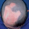
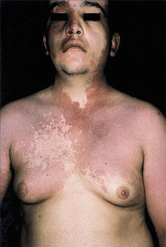
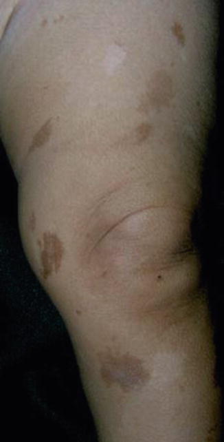
A hallmark of this disorder is the presence of rather large light and dark macules on a background skin of intermediate color (Fig. 8.3), sometimes being arranged in a distinct sash-like pattern (Fig. 5.26) (see Sect. 5.6). Extracutaneous mosaic defects involve the brain [10, 17, 20] and the bones [21].
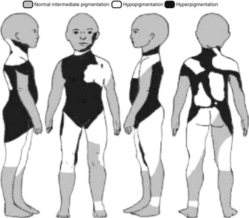
Fig. 8.3
Cutis tricolor arranged in large patches without midline separation as noted in the Ruggieri-Happle syndrome [20]
8.1.2.2 Cutis Tricolor Parvimaculata
8.1.2.3 Cutis Tricolor of the Blaschko-Linear Type
Niessen et al. [18] described hyper- and hypopigmented bands following Blaschko’s lines in a 6-year-old girl with double aneuploidy mosaicism 47,XX+7/45,X.
8.1.3 Didymosis in Epidermolytic Ichthyosis of Brocq
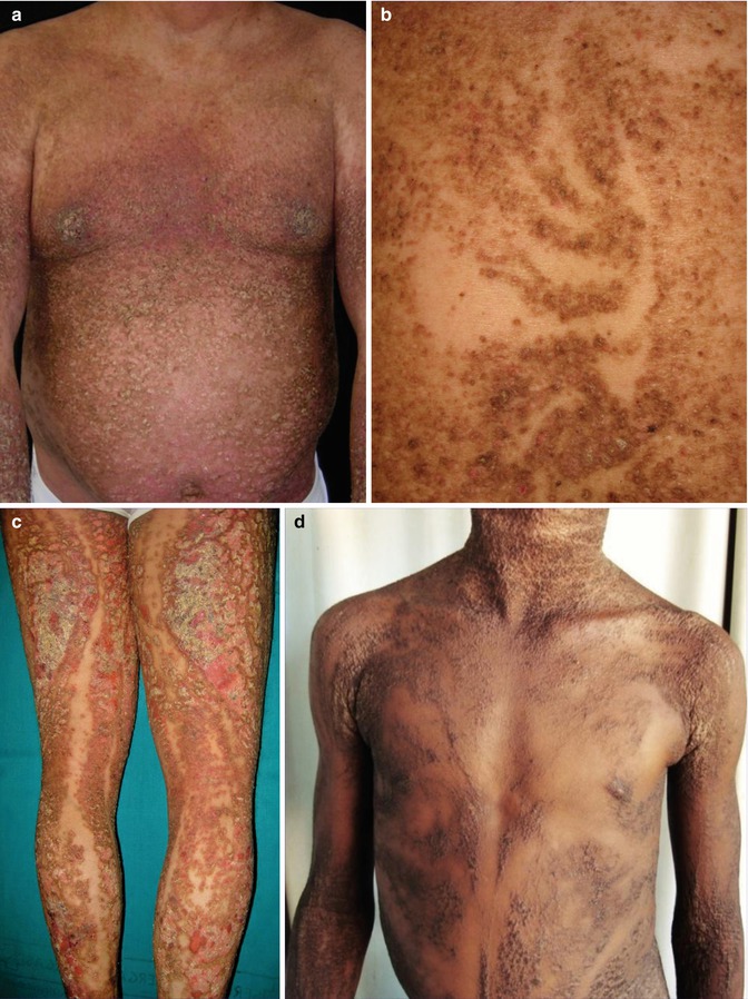
8.1.4 Didymosis in Darier Disease
Patients with Darier disease may likewise have paired bands of either pronounced or absent involvement, which can be interpreted as cases of allelic twin spotting (Fig. 8.5). Three clinical examples suggestive of such mechanism have so far been reported [14, 19, 26], but molecular evidence is still lacking.