Diagnosing nail matrix diseases requires knowledge of the nail matrix function and anatomy. This allows recognition of the clinical manifestations and assessment of potential surgical risk. Nail signs depend on the location within the matrix (proximal or distal) and the intensity, duration, and extent of the insult. Proximal matrix involvement includes nail surface irregularities (longitudinal lines, transverse lines, roughness of the nail surface, pitting, and superficial brittleness), whereas distal matrix insult induces longitudinal or transverse chromonychia. Clinical signs are described and their main causes are listed to enable readers to diagnose matrix disease from the nail’s clinical features.
Key points
- •
The proximal portion of the matrix produces the upper third of the nail plate and its distal part produces the lower two-thirds.
- •
Insult to the proximal matrix produces nail surface irregularities: longitudinal lines, transverse lines, roughness of the nail surface, pitting, or superficial brittleness.
- •
Surgery on the distal matrix is at very low risk of inducing nail dystrophy, because the upper layers of the nail plate cover the defect.
- •
Involvement of the distal matrix presents as a longitudinal, transverse, or diffuse chromonychia (melanonychia, erythronychia, leukonychia).
- •
The growth rate of the plate is 3 times less on a toenail (1 mm/mo) than on a fingernail (3 mm/mo), which allows clinicians to date retrospectively an event that caused nail surface dystrophy.
The first question to be answered when facing a nail dystrophy is: where is the primary seat of the disease? Clinical nail examination allows identification of the affected component of the nail apparatus. In order to fully understand how a nail matrix disease may manifest clinically, it is mandatory to know its physiologic function and anatomy.
Gross nail matrix anatomy
The nail matrix covers the bottom of the cul-de-sac and rises on the posterior quarter (or even less) of the ventral aspect of the proximal nail fold. The matrix rests on the base of the distal bony phalanx and forms a crescent with posterior inferior concavity ( Fig. 1 A). Clinicians should bear in mind that, on the great toes, both lateral ends of the crescent (also called the lateral horns of the matrix) expand much more proximally on the lateral aspect of the phalanx than on the fingers (see Fig. 1 B). The lateral horns may reach to or even beyond the midline of the lateral aspect of the great toe. This anatomic feature explains why spicules are the most common complication of surgical treatment of ingrown toenail and lateral longitudinal biopsies by unskilled clinicians.
Nail matrix histology
- 1.
The nail matrix has a multilayered epithelium. Keratinization occurs without formation of keratohyaline granules. It gives rise to the nail plate. In the midline of the nail unit, the matrix epithelium is thick with long, oblique, distally oriented rete ridges. The nail matrix epithelium is the sole site of hard keratin synthesis.
- 2.
Nail matrix melanocytes are 6 times less numerous in the matrix (200/mm 2 ) than in the skin epidermis (1200/mm 2 ). Their numbers are similar in the proximal and the distal matrix, but they differ in their usual quiescence: in the proximal matrix, most matrix melanocytes are dormant and do not produce any pigment whereas in the distal matrix 50% are dormant and 50% are activable. When activated, they synthesize melanin, which is transferred to the surrounding keratinocytes. Distal migration of melanin-containing keratinocytes gives rise to a pigmented nail plate. For this reason, most longitudinal melanonychias arise in the distal matrix, which is fortunate if they have to be surgically removed (discussed later).
Formation of the nail plate
- 1.
The matrix creates all or most of the nail plate. The proximal portion of the matrix produces the upper third of the nail plate and its distal part produces the lower two-thirds (see Fig. 1 C). This feature is important in nail surgery: removing a part of the distal matrix (eg, with a punch) does not lead to nail dystrophy because the defect will be covered by the upper part of the plate synthesized by the proximal matrix (see Fig. 1 D).
- 2.
The thickness of the nail plate is proportional to the length of the matrix (thumbnails and great toenails are thicker).
- 3.
The shape of the lunula determines the contour of the free edge.
Growth of the nail plate
The average fingernail growth rate (3.47 mm/mo) is more than twice as fast as that of the toenail (1.62 mm/mo). This rate has increased over the last 30 years thanks to the improvement in quality of life, health conditions, and diet. This growth rate allows clinicians to date an event that left a nail surface dystrophy (eg, Beau line, hematoma) by measuring the distance between the cuticle and the dystrophy.
Clinical signs of nail matrix insults
The resultant nail signs depend on the intensity (mild, moderate, or severe), the duration (transient or prolonged), and the extent (focal, widespread) of the insult.
Because the proximal matrix synthesizes the upper part of the nail plate, an insult to the proximal matrix produces nail surface irregularities: longitudinal lines, transverse lines, roughness of the nail surface, pitting, or superficial brittleness.
When the distal matrix is affected, the condition may present as a longitudinal, transverse, or diffuse chromonychia (melanonychia, erythronychia, leukonychia).
Clinical signs of proximal matrix involvement
Longitudinal Lines May Appear as Projecting Ridges or Indented Grooves
Longitudinal ridging
Multiple shallow and delicate longitudinal ridging is physiologic. This relief becomes more prominent with aging ( Fig. 2 ). The ridges may be focally interrupted, giving rise to a beaded appearance. In contrast with what is noted in young adults, there is a discrepancy and an irregularity in the turnover of the matrix cells. These variations are probably responsible for the exaggeration of the longitudinal ridges observed on senile nails. Ridges may also be observed in some pathologic states, such as rheumatoid arthritis, peripheral vascular disorders, and Darier disease.
Sometimes both thumbs have a wide median longitudinal ridge that has the shape of a circumflex accent in cross section. This condition may be inherited or, if acquired, is posttraumatic.
Longitudinal grooves
These depressions may run on a part or on the whole nail length. They can be single, multiple, on 1 or several nails, and are associated with a pathologic state in almost all instances. They arise from a focal prolonged moderate insult to the proximal matrix. Depending on the depth, length, and width of the depression, clinicians talk about fissures (narrow and superficial), cracks (short, deep and narrow), splits (long, deep, and narrow), grooves (wide, long and deep), or gutters (larger grooves).
Onychorrhexis defines multiple superficial fissures that give the appearance of the nail having been scratched with coarse sandpaper or with an awl. Some splits may be associated. These fissures are the most common presentation of lichen planus ( Fig. 3 ).
The median canaliform dystrophy of Heller is characterized by a midline inverted fir tree–like crack emerging from under the cuticle and extending up to two-thirds of the proximal nail plate ( Fig. 4 ). It rarely reaches the free edge. This dystrophy electively affects the thumbnails. Its cause remains unclear and it is probably a self-inflicted repeated pressure on the proximal region of the nail, as indicated by the enlarged lunula and its disappearance with protection of the proximal nail. Two cases have been attributed to isotretinoin, both improving on withdrawal of the drug.
A single smooth and harmonious longitudinal gutter results from prolonged focal pressure on the underlying matrix by a tumor located on or under the proximal nail fold. These tumors are mainly benign: myxoid pseudocyst ( Fig. 5 ), fibrokeratoma ( Fig. 6 ), implantation cyst, wart, and giant-cell tumor of the tendon sheath. Myxoid pseudocysts usually imprint a smooth wide gutter on the plate. However, some may vary in size: inflammation or drainage temporarily lessens their pressure on the matrix before they swell again. This process results in a typical irregular groove of varying depths ( Fig. 7 ). No other tumor varies in size. The main causes of longitudinal lines are listed in Table 1 .
| Single median fissure | Trauma Median canaliform dystrophy of Heller |
| Gutter | Tumor of the proximal nail fold |
| Onychorrhexis | Lichen planus Rheumatoid arthritis Raynaud disease |
| Multiple longitudinal ridges | Physiologic Old age Rheumatoid arthritis |
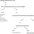
Transverse Lines Are Always Grooves
Transverse grooves originating from the matrix always parallel the distal edge of the lunula. Transverse grooves following the curve of the proximal nail fold have an exogenous origin (eg, manicuring) ( Fig. 8 ). The French physician Joseph Honoré Simon Beau first described this nail dystrophy in 1846 after typhoid fever and other acute systemic diseases. Beau was a cardiologist, but the medical community remembers him for noticing this condition. The groove results from a transient decrease of the matrix mitotic activity, resulting in a focal thinning of the nail plate. The groove is more pronounced in the middle part of the nail. Beau lines emerge from under the cuticle 4 to 8 weeks after the matrix insult ; they grow out distally with the nail growth. Because the nail plate grows at a speed of 0.10 mm/d, it is easy retrospectively to estimate the timing of the causal event by measuring the distance between the groove and the proximal nail fold. One single transverse groove on 1 nail is secondary to a local phenomenon (eg, trauma) ( Fig. 9 ). The existence of 1 groove on several nails, at the same level, reflects a systemic event. Several Beau lines on all nails suggest a repeated insult to the matrix (eg, chemotherapy) ( Fig. 10 ). Neonates may show Beau lines at 8 to 9 weeks of age, reflecting the transition from intrauterine to extrauterine life. The main causes for Beau lines are listed in Table 2 .
| Polydactylous single groove | Birth Infectious disease Feverish disease |
| Monodactylous single groove | Trauma Surgery Acute paronychia Carpal tunnel syndrome |
| Monodactylous multiple grooves | Habit tic deformity (thumbs almost exclusively) Chronic paronychia |
| Polydactylous multiple grooves | Regular cosmetic traumatism Retinoids Eczema Successive chemotherapies |
Onychomadesis refers to a proximal detachment of the plate followed by nail shedding. It appears after an insult to the proximal matrix that lasts for more than 1 or 2 weeks or after a complete arrest of the matrix activity. A new nail is formed and slides under the former nail, which is lifted up and pushed away. Onychomadesis has gain popularity lasting recent years from epidemic outbreaks of hand-foot-and-mouth syndrome associated with this nail dystrophy ( Fig. 11 ).
Multiple parallel transverse grooves, especially on the thumbnails, spreading from the cuticle up to the free edge, located in the middle of the plate and giving rise to a characteristic washboard aspect, indicate a self-induced dystrophy. This condition is called habit tic and the dystrophy comes from the repeated pushing back of the cuticle from the thumb with the nail of the index or middle finger of the same hand or the contralateral thumbnail. This habit leads to transient repeated widespread insult to the matrix, resulting each time in a moderately deep transverse groove. If the habit exerts a sufficient and constant strong pressure on the proximal area, it also leads to a wide longitudinal groove where the transverse grooves rest ( Figs. 12 and 13 ). Pressure induces an enlargement of the lunula (macrolunula), which is an excellent diagnostic clue. Cuticles may be partially or totally absent and a subacute paronychia may be associated. A habit tic was recently associated with guitar playing, and in one case it was responsible for an added elkonyxis.