53
Developmental Anomalies
Developmental anomalies are a diverse group of congenital disorders that result from faulty in utero morphogenesis. When they affect the skin, developmental anomalies can range in severity from isolated minor physical findings to potentially life-threatening conditions or cutaneous signs of significant extracutaneous defects.
Midline Lesions of the Nose or Scalp
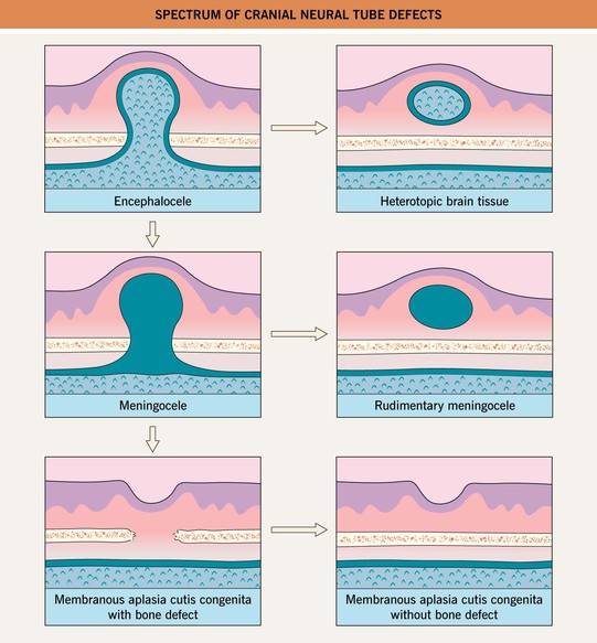
• A midline mass or pit on the nose or scalp due to a dermoid cyst, cephalocele, nasal glioma, or other heterotopic brain/meningeal tissue (Fig. 53.1) may have a deeper component with intracranial extension.
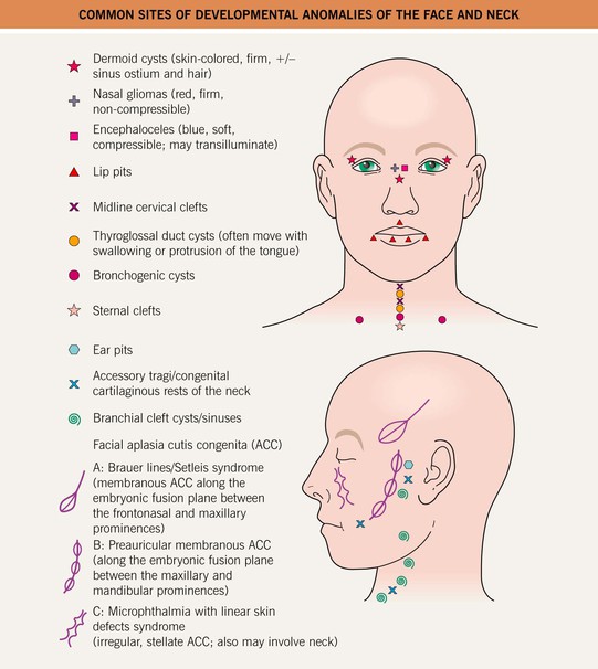
Fig. 53.1 Common sites of developmental anomalies of the face and neck. Courtesy, Julie V. Schaffer, MD.
• These lesions are typically apparent at birth or during early childhood.
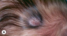
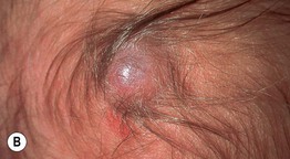
• Hair collar sign: a peripheral ring of long, dark hair often surrounds ectopic neural tissue or membranous aplasia cutis congenita (ACC) on the scalp (Figs. 53.2 and 53.3); the latter is thought to represent a forme fruste of a neural tube defect.
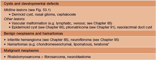
• DDx: outlined in Table 53.1 for nasal masses; epidermoid and pilar cysts for scalp masses (especially in older children and adults).
Dermoid Cysts
• Result from sequestration of ectodermal tissue along embryonic fusion planes.
• Recognized at birth or when they enlarge or become inflamed during infancy or childhood.
• Most often located around the eyes, especially the lateral eyebrow region (Fig. 53.4); midline lesions on the nose, scalp, or back may have intracranial extension.
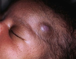
Fig. 53.4 Dermoid cyst. This dermoid cyst presented in an infant as a firm subcutaneous nodule superior to the lateral left eyebrow. Courtesy, Kalman Watsky, MD.
Cephaloceles
Nasal Gliomas, Other Heterotopic Brain Tissue, and Rudimentary Meningoceles
• A nasal glioma is a congenital mass of heterotopic brain tissue (HBT) at the nasal root/glabella > intranasally; the skin overlying this firm, noncompressible nodule tends to be red with prominent telangiectasias (mimicking an infantile hemangioma) (Fig. 53.5).
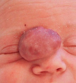
Fig. 53.5 Nasal glioma. A mass was noted on prenatal ultrasound, and this reddish, slightly pedunculated, rubbery nodule was evident at birth. Courtesy, Mary Chang, MD.
• Other HBT and rudimentary meningoceles typically present as a solid or cystic subcutaneous nodule on the midline scalp, often with a blue-red hue, overlying alopecia and a surrounding hair collar (see above); a rudimentary meningocele may have a bullous appearance and can also be located over the spine.
• May have a vestigial fibrous stalk extending into the intracranial space, but lack a connection with the intracranial leptomeninges or CSF (see Fig. 53.3).
Midline Cervical, Sternal, and Supraumbilical Clefts
• A sternal cleft or supraumbilical raphe presents with a band of atrophic (Fig. 53.6), scarred or ulcerated skin; may occur in the setting of PHACE(S) syndrome (posterior fossa malformations; hemangiomas; arterial, cardiac, and eye anomalies; sternal cleft/supraumbilical raphe; see Chapter 85).
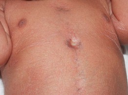
Fig. 53.6 Sternal cleft. Note the atrophic skin overlying the defect and prominent veins in the midline chest. Generalized desquamation is also evident in this 1-day-old post-term neonate. She did not develop an infantile hemangioma and had no cardiac defects or other features of PHACE(S) syndrome. Courtesy, Julie V. Schaffer, MD.
Midline Lesions Overlying the Spine
• Skin lesions associated with spinal dysraphism are presented in Table 53.2 and Fig. 53.7.
Table 53.2
Skin lesions of the spinal axis associated with dysraphism.
The presence of two or more types of lesions increases the risk of a spinal anomaly.
| Lesion | Features |
| Hypertrichosis | A V-shaped patch of long, coarse or silky hair (see Fig. 53.7A,B); ‘faun tail’ |
| Lipomas | Soft subcutaneous mass, asymmetric buttocks, curved gluteal cleft (see Fig. 53.7C); most common sign of spinal dysraphism |
