Fig. 22.1
Retention of perifollicular pigmentation in early lesion of vitiligo
22.4 Dermoscopic Findings in the Diagnosis of Early Vitiligo
Thatte and Khopkar [3] in their recent study have reported the following dermoscopic findings in evolving vitiligo: reduced/absent (Fig. 22.2), reversed pigmentary network (Fig. 22.3), perifollicular and perilesional hyperpigmentation (Fig. 22.4), and a diffuse white glow under UV light. The predominant findings noted were reduced (40 %), absent (30 %), and reversed pigmentary network (20 %).
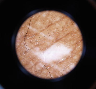
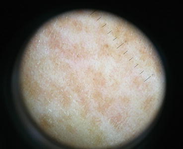
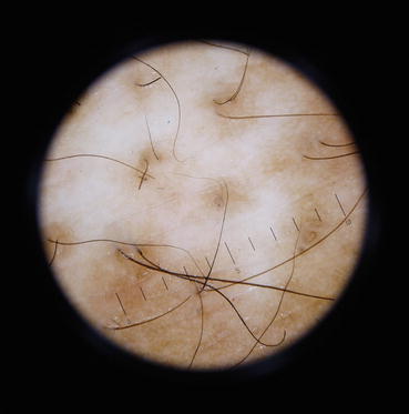
Fig. 22.2
Absence of pigment network with Koebner phenomenon in an early lesion of vitiligo
Fig. 22.3
Reversed pigmentary network in an early lesion of vitiligo
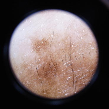
Fig. 22.4
Perifollicular pigmentation with reduced pigment network in an early lesion of vitiligo
Reversed pigmentary network, a well-described finding in the dermoscopy of melanoma and melanocytic nevus, refers to a white or depigmented net-like pattern with pigmentation. It is similar to the salt and pepper pattern reported by Chandrashekar [2]. In vitiligo, there is a gradual loss of melanocytes and melanin which allows the light to directly pass into the dermis without being reflected by the melanocytes and melanin. This leads to a window through which light passes into the dermis and is reflected by dermal collagen.
In the initial stages of evolving vitiligo, this leads to an area of relative hyperpigmentation produced by the pale area corresponding to the papillary dermis in the normal reticulate pattern of pigmentation. The appearance of “reversed pigmentary network pattern” in evolving vitiligo is thus seen.
Identification of leukotrichia in nonlesional skin on dermoscopy may also be considered as an earlier dermoscopic marker of impending vitiligo. In a recent report by Sonthalia et al. [8], perifollicular depigmentation and evolving leukotrichia in areas of clinically unaltered pigmentation has been suggested as an early predictive sign of impending vitiligo.
22.5 Dermoscopic Features to Assess the Stage of Vitiligo
Assessing the stability of vitiligo when considering vitiligo surgery is another area where the dermoscope comes as a handy tool. There are various well-described clinical signs of progressive activity like confetti macules, koebnerization, onset of new macules, and trichrome. In a Chinese study [9], it was reported that residual perifollicular pigmentation was observed in 91.94 % of patients with progressive vitiligo when compared to 62.86 % of those with stable vitiligo. There was no residual perifollicular pigmentation observed in nonvitiligo depigmented lesions. The presence of telangiectasia, early reservoirs of pigmentation, and perilesional hyperpigmentation was reported to be related to the stage of vitiligo and treatment history of patients.
Chandrashekar [2] has reported various signs associated with stable and progressive vitiligo. The various dermoscopic findings associated with stability and repigmentation of vitiligo are marginal and perifollicular hyperpigmentation (Fig. 22.5), reticular pigmentation (well-defined pigment network within the depigmented macule), and marginal reticular pigmentation (well-defined pigment network at the margins of the macule) (Fig. 22.6). The various dermoscopic findings associated with progressive vitiligo include polka dot or confetti-like (depigmented dots distributed in a polka dot pattern) (Fig. 22.7), comet tail (micro-Koebner phenomenon) (Fig. 22.8), starburst or nebulous (Fig. 22.9), trichrome vitiligo (three zones, brown, tan, and white) (Fig. 22.10), and salt and pepper pattern.
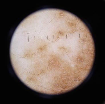
Fig. 22.5
Perifollicular hyperpigmentation in a case of stable repigmenting vitiligo