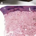
Histopathology:
Dense dermal infiltrate of neutrophils, sometimes with papillary dermal edema (Fig. 11.1C)
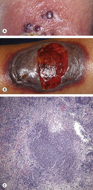
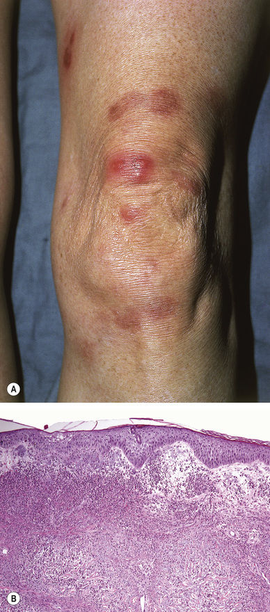
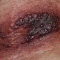
Erythema Elevatum Diutinum, Acute Stage (Fig. 11.3)
Symmetric, often acral (extensor surfaces of elbows/hands), red–violet to pink–brown papules and plaques
Histopathology:
Infiltrate of neutrophils with vascular damage
Late stage becomes clinically indurated and histologically fibrotic (see Fig. 22.16)
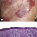
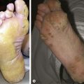
Cellulitis (Fig. 11.4)
Any site, but commonly affects the lower legs
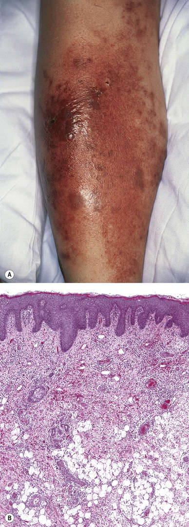
Fig. 11.4 Cellulitis (group A streptococci). A, Courtesy, Yale Dermatology Residents’ Slide Collection. A, From Bolognia JL, Jorizzo JL, Schaffer JV. Dermatology, 3e. London: Saunders, 2012, with permission. B, From Guarner J. Skin and soft tissue infections. In: Procop GW, Pritt BS (eds). Pathology of Infectious Diseases. London: Saunders, 2015.
Warm, tender, bright red plaque
Often associated fever and elevated white cell count
Histopathology:
Interstitial infiltrate of neutrophils
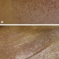
Mixed With Neutrophils and Eosinophils or Predominant Eosinophils (Pink–Red)
Wells’ Syndrome (Eosinophilic Cellulitis; Fig. 11.5)
Pink edematous plaques that may resemble cellulitis
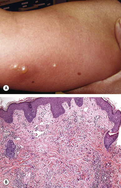
Histopathology:
Interstitial infiltrate of eosinophils and neutrophils, often with flame figures (collagen encrusted with granular red–purple material)
Related posts:

Stay updated, free articles. Join our Telegram channel

Full access? Get Clinical Tree


