81
Dermal Hypertrophies
Hypertrophic Scar
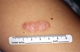
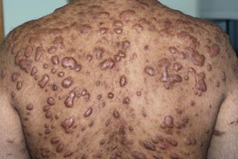
• Firm, initially pink to purple in color then becomes skin-colored to hypopigmented, occasionally hyperpigmented; papule or plaque limited to an excision site or wound (Figs. 81.1 and 81.2).

Fig. 81.1 Hypertrophic scar at the site of an excision of an atypical melanocytic nevus. The deltoid region is a common location for hypertrophic scars. Courtesy, Jean L. Bolognia, MD.
• Most commonly seen on the trunk/shoulders.
• With treatment, can reduce pruritus and height but not width of scar.
Keloid
• More common in patients with darkly pigmented skin who have a familial predisposition.
• Rarely associated with syndromes (e.g. Rubinstein–Taybi or Goeminne syndromes).
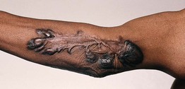
• Raised, often skin-colored firm plaque(s) that, in contrast to hypertrophic scars, extend beyond the wound margin (Fig. 81.3; Table 81.1); color may vary as in hypertrophic scars.


