What are the different types of teeth that are present in the adult dentition?
1. Incisors—anterior teeth, single rooted, used for biting/incising
2. Canines—anterior teeth, cornerstones of the mouth, prominent cusp tips, single long roots
3. Premolars—aka bicuspids, posterior teeth, occlusal surface to chew, commonly single rooted in mandible and dual rooted in maxilla
4. Molars—posterior teeth used primarily for chewing, maxillary molars with three roots and mandibular molars with two roots
See Figure 54-1.
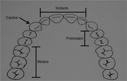
Figure 54-1 Types of teeth.
 You are called in to consult on a 27-year-old male who has sustained facial trauma. He has multiple intraoral and perioral lacerations and the crowns of his two maxillary central incisors are no longer present in his mouth. When calling the dental trauma consultant, which teeth will you tell her are fractured?
Teeth 8 and 9.
The Universal Numbering System has been adopted by the American Dental Association. NUMBERS designate the 32 permanent (adult) teeth (1–32). Begin at the RIGHT maxillary third molar (#1) to the LEFT maxillary third molar (#16), down to the LEFT mandibular third molar (#17), and end at the RIGHT mandibular third molar (#32). Many individuals will have impacted third molars or have had their third molars extracted. Therefore, you will frequently begin counting at tooth #2.
LETTERS designate the 20 primary (deciduous) teeth (A–T). See Figure 54-2.
Other systems in use include the International Numbering System and the Palmer Notation Method.
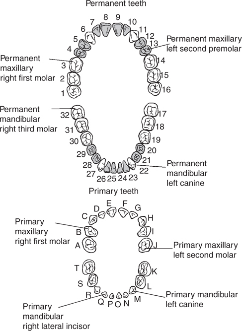
Figure 54-2 Universal numbering. Reproduced with permission from Tintinalli JE, Kelen GD, Stapczynski JS, et al. Tintinalli’s Emergency Medicine: A Comprehensive Study Guide. 6th ed. New York, NY: McGraw-Hill; 2004. Fig. 242–2.
 What are the parts of the tooth and its housing?
1. Enamel—hard outer highly mineralized covering of the tooth crown composed mainly of hydroxyapatite.
2. Dentin—less mineralized structure than enamel that also includes organic material and water containing microtubules. When exposed, teeth become sensitive.
3. Cementum—Located only on the root, it covers the dentin. It is less calcified than enamel or dentin and attaches to the periodontal ligament.
4. Pulp—neurovascular component of teeth also containing connective tissue and cells (odontoblasts). Pulpal inflammation or exposure causes significant pain.
5. Periodontal ligament—connective tissue that attaches cementum to bone, allowing for movement of the tooth in the socket as well as proprioception.
6. Alveolar bone—made of cortical and cancellous bone.
See Figure 54-3.
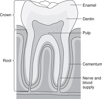
Figure 54-3 Anatomy of the tooth and surrounding structures. Reproduced from Stone CK, Humphries RL. Current Diagnosis & Treatment: Emergency Medicine, 2008. 6th ed. New York, NY: McGraw-Hill. Fig. 30–11.
 Describe the following anatomic terms used in describing surfaces of teeth:
1. FACIAL: part of tooth that faces the lips or cheeks. Can be further subdivided into:
a. LABIAL: facial surface of ANTERIOR teeth (incisors and canines).
b. BUCCAL: facial surface of POSTERIOR teeth (premolars and molars).
2. PALATAL: surface of maxillary teeth that face the palate.
3. LINGUAL: describes both maxillary and mandibular surfaces that face the tongue.
4. MESIAL: TOWARD the dental midline.
5. DISTAL: AWAY from the dental midline.
6. OCCLUSAL: the chewing surface of the POSTERIOR teeth.
7. INCISAL: the biting surface of the ANTERIOR teeth.
See Figure 54-4.
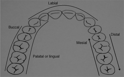
Figure 54-4 Surfaces of teeth.
 Describe the following dental terminology.
CORONAL: refers to the crown of the tooth, which is the portion of the tooth which is visible in the mouth. “Coronal” can be used as a directional term. For example, the incisal edge of an incisor is coronal to the gingiva.
APICAL: the apex of the tooth is at the root end. The root is the portion of the tooth which is housed in the alveolar bone. The term “apical” can also be used as a directional term. For example, starting at the incisal edge and moving apically, one would reach the root of the tooth.
CINGULUM: the cingulum is a bulge on the lingual surface of anterior teeth.
Related posts:
Stay updated, free articles. Join our Telegram channel

Full access? Get Clinical Tree


