34
Delayed Treatment of Flexor Tendons: Staged Tendon Reconstruction
Lawrence H. Schneider
History and Clinical Presentation
An 18-year-old student lacerated both flexor tendons in zone 2 of his right dominant index finger on broken glass. His primary treatment was by direct repair of both flexor tendons 3 days postinjury, and he was started on a mobilization program in a hand therapy unit. Unfortunately, this did not result in functional pull-through of his flexor tendons. He then underwent flexor tenolysis in an attempt to salvage function, a procedure that was also not successful. He was first seen on my hand surgery service at 4 months postoperative complaining of restricted motion in the operated finger.
PEARLS
- The patient must demonstrate an ability to comply with a detailed rehabilitative program.
- Staged reconstructions are not a perfect technique, but they are the only way to restore active motion at the interphalangeal joints of the finger in these difficult cases of flexor system disruption.
- Never hesitate to build pulleys! Pulleys need to be strong. Although there are many techniques for pulley reconstruction, I prefer to wrap the tendon material around the proximal phalanx at A2 as was done in this case. At A4 I have most frequently used local tissue sutured over the implant, often with the addition of tendon material.
PITFALLS
- Complications are not infrequent.
- This is not a simple procedure for patient or surgeon.
- Severe flexion contractures especially after stage 1 are poor prognostic signs.
- Double digital nerve injury is a very poor preoperative sign. When present, it may be an indication for amputation or fusion (if the finger is in a nonfunctional position).
Physical Examination
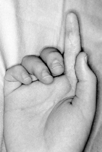
The right hand showed no active flexor tendon function at either the flexor digitorum superficialis (FDS) or flexor digitorum profundus (FDP) of the right index finger (Fig. 34–1). Passive motion was almost full with only a mild flexion deformity at the proximal interphalangeal (PIP) joint. Sensory nerve function was intact. The skin was relatively pliable and soft considering that he had had an injury and two prior operations. The remainder of the hand was normal.

Figure 34–1. Right index finger held in extended position while attempted to make a full fist even after two operative procedures.
Diagnostic Studies
Except for routine radiographs in cases with associated bone injury or arthritis, there are no diagnostic studies needed. Although there is some interest in the use of ultrasound and magnetic resonance imaging (MRI) in the evaluation of the flexor tendon system, I have not used these studies for these cases.
Diagnosis
Flexor System Laceration of Right Index Finger, Status Postoperative Repair, and Tenolysis with Loss of Flexor Tendon Function
Stay updated, free articles. Join our Telegram channel

Full access? Get Clinical Tree


