Darker skin types

21 Darker skin types


Racial / ethnic differences in photoaging


Facial rhytides are a common concern across the racial and ethnic spectrum. However, variations in severity, age of onset, and cultural impact can be seen in different populations. In general, as the studies by Hexsel & Brunetto and Rossi & Alexis reported, individuals with richly pigmented skin (i.e. Fitzpatrick skin types IV–VI) demonstrate signs of photoaging, including rhytides, at a later age than do individuals with fair skin (Fitzpatrick skin types I–III). This is largely due to the photoprotective effect of increased epidermal melanin, which has been demonstrated in several studies. Most notably, the mean protective factor from ultraviolet (UV) B in black skin was found to be 13.4 versus 3.4 for white skin in a cadaveric skin study by Kaidbey and colleagues. Furthermore, in a more recent study by Yamaguchi and co-workers, UV-induced apoptosis was found to be greater in the epidermis of black skin compared with white skin, suggesting that photodamaged cells may be removed more efficiently in darker phototypes.


In a comparative study by Nouveau-Richard et al of Chinese and French women, the onset of facial wrinkles was found to be approximately 10 years later in Chinese versus French women. Other studies in African-Americans have reported similar findings of later onset of facial rhytides or fine lines; Grimes found a much lower percentage of women of color perceived having wrinkles than did their Caucasian counterparts. In general, signs of facial aging in darker skin occur 10–20 years later than in Caucasians, and is more often evident in the deeper muscular layers in the face.


Loss of volume in the mid-face and prominent tear troughs have also been reported to be more striking features of aging in African-Americans. Perioral rhytides are uncommon in darker skin types and therefore the upper face is the primary site of facial rhytides in patients with skin of color. Photoaging differences in Hispanics / Latinos are less well characterized, but vary considerably, given the broad range of Fitzpatrick skin types (I–VI) found in this population.


Safety and efficacy of botulinum toxins in darker skin types


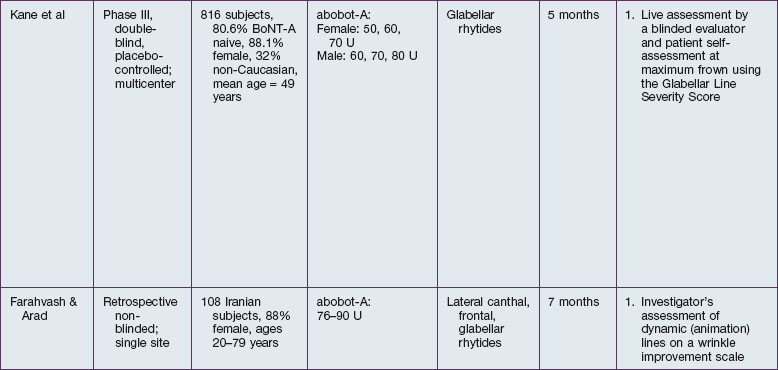
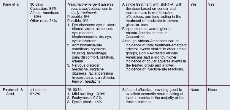
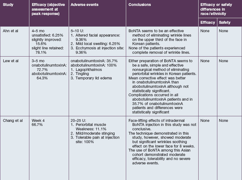
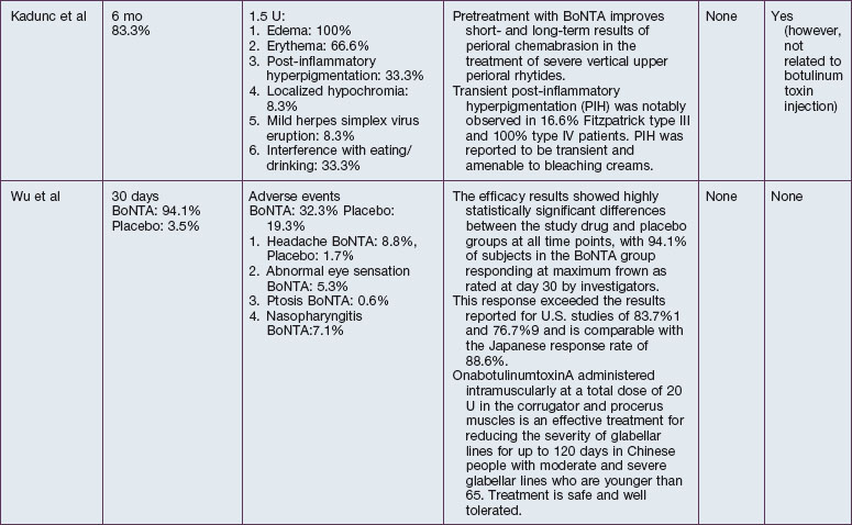
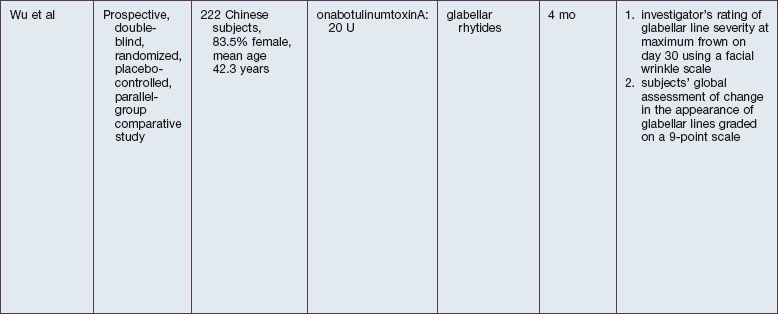
The safety and efficacy of botulinum neurotoxin type A in the treatment of glabellar lines has been well studied in numerous populations (Table 21.1). Published data pertaining to the safety and efficacy of botulinum toxin in non-white patient populations are reviewed herein.


A multicenter, double-blind, placebo controlled study of onabotulinumtoxinA (onabot-A) (Botox Cosmetic®, Allergan, Inc., Irvine, CA) by Carruthers and colleagues in 2002 investigated safety and efficacy in the treatment of glabellar lines in 409 patients. No appreciable differences were observed between various skin phototypes.


African-Americans


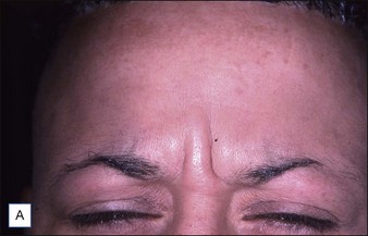
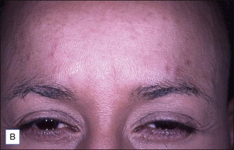
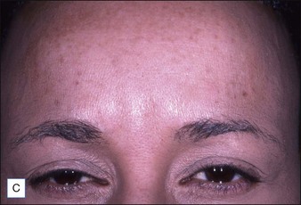
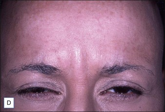
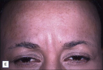
Grimes & Shabazz conducted a Phase IV study of onabot-A in the treatment of glabellar lines in 31 African-American women with Fitzpatrick skin types V and VI. The authors assessed the safety and efficacy at doses of 20 and 30 units (U) of onabot-A. No statistically significant differences in efficacy or safety were observed between the two doses (Figs 21.1, 21.2). A maximal response was observed on day 30, with 92.4% and 100% response rates (i.e. a score of ‘none’ or ‘mild’ on the facial wrinkle scale) in the 20 U and 30 U groups respectively. Adverse events were mild and transient and did not differ between the dosing groups. They included mild tingling, slight headaches, and dullness of the forehead.


Stay updated, free articles. Join our Telegram channel

Mar 12, 2016 | Posted by in General Surgery | Comments Off on Darker skin types

Full access? Get Clinical Tree
Get Clinical Tree app for offline access