90
Cysts
Introduction
True Cysts – Common
Epidermoid Inclusion Cyst (EIC) (Epidermal Inclusion Cyst, ‘Sebaceous Cyst’)
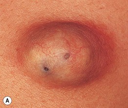
• A visible comedonal-like opening or pore (resembles a blackhead) may be seen on the surface of the papule or nodule (Fig. 90.1A).


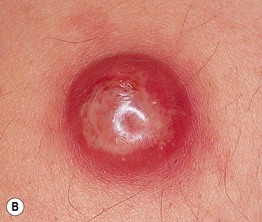
Fig. 90.1 Epidermoid inclusion cysts. A Typical clinical appearance of an epidermoid inclusion cyst with a yellowish hue. Two pores are present in this example. B Painful inflammatory reactions to cyst rupture are a frequent cause for presentation to a physician. Culture will often prove these lesions to be sterile, even if draining pus. Antibiotic treatment can be helpful due to their anti-inflammatory effects. B, Courtesy, Mary S. Stone, MD.
• Cyst contents may rupture into the dermis, eliciting an acute and chronic inflammatory reaction, leading to significant redness and pain; this inflammation is often confused with a bacterial infection (Fig. 90.1B).
• Most commonly sporadic; multiple lesions rarely associated with Gardner syndrome or Gorlin syndrome.
Milium (Milia – Plural)
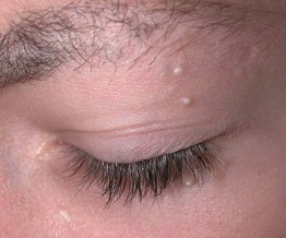
• A small, superficial (1–2 mm), firm cyst that is white in color and is sometimes confused with a whitehead (Fig. 90.2); occasionally they are grouped.
• Commonly observed on the face in newborns; in this setting, they often resolve spontaneously.
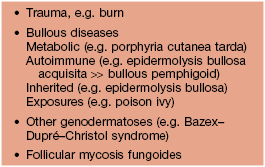
• The majority of patients with multiple facial milia have no underlying condition; however, there may be a secondary cause (Table 90.1).
• DDx:
Stay updated, free articles. Join our Telegram channel

Full access? Get Clinical Tree


