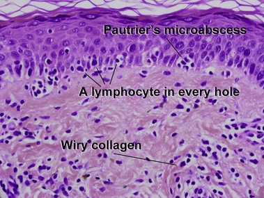
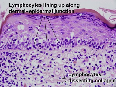
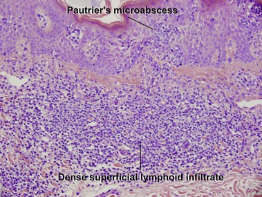
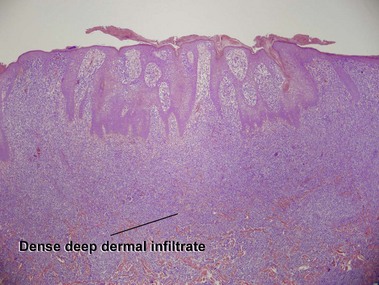
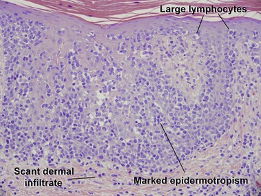
Chapter 24 Table 24-1 Primary cutaneous T-cell lymphomas, NK-cell lymphomas, and precursor hematologic neoplasm in the 2008 WHO classification Mature T-cell and NK-cell neoplasms Mycosis fungoides variants and subtypes Adult T-cell leukemia/lymphoma Primary cutaneous CD30+ T-cell lymphoproliferative disorders Subcutaneous panniculitis-like T-cell lymphoma Extranodal NK/T-cell lymphoma, nasal type Primary cutaneous peripheral T-cell lymphoma, rare subtypes Primary cutaneous aggressive epidermotropic CD8+ cytotoxic T-cell lymphomaa Primary cutaneous gamma–delta T-cell lymphoma Precursor hematologic neoplasm Adapted from WHO Classification of Tumours of Haematopoietic and Lymphoid Tissues, 4th edn. Lyon, France: IARC; 2008. Mycosis fungoides (MF) is the most common type of cutaneous lymphoma. In most cases, the disease is indolent and slowly progressive over a period of years or decades. Three main stages of the lymphoma are recognized: patch, plaque, and tumor stages. In the patch stage of mycosis fungoides, patients typically present with broad pink or tan oval-shaped patches with a predilection for the bathing trunk area. The patches may be asymptomatic or pruritic. Both clinically and histopathologically, distinction from eczematous dermatitis is sometimes difficult in the earliest stages of the lymphoma. In the evaluation of patch stage mycosis fungoides, multiple shave biopsies are often helpful, since the shave technique provides a broad area of epidermis for examination. The typical immunophenotype is CD3+, CD4+, CD8−, CD30−. Aberrant immunophenotypes (with loss of normal T-cell markers, such as CD7) can frequently be demonstrated. Clonal rearrangement of the T-cell receptor gene is helpful in supporting the diagnosis, although the earliest cases may sometimes not have a detectable clone.
Cutaneous T-cell lymphoma, NK-cell lymphoma, and myeloid leukemia
Cutaneous T-cell lymphoma and NK-cell lymphoma
Mycosis fungoides
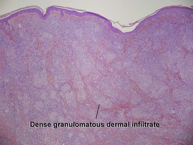
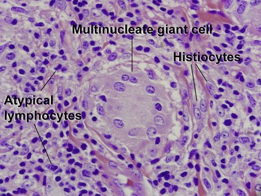
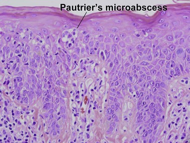
Patch stage
Cutaneous T-cell lymphoma, NK-cell lymphoma, and myeloid leukemia