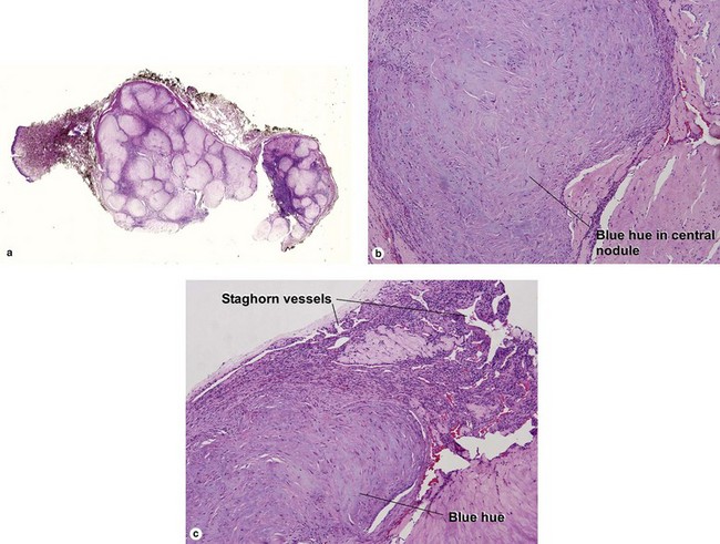
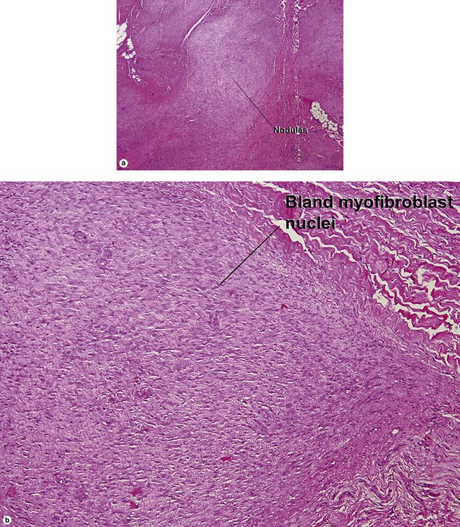
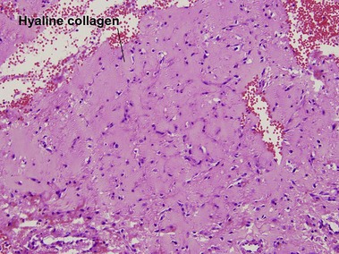
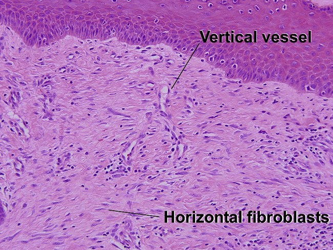
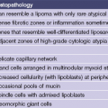
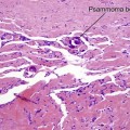
Chapter 20 The shade of blue in the center of the nodule resembles that of cartilage. The peripheral vascular proliferation may have staghorn vessels and resemble hemangiopericytoma. There is a tendency towards spontaneous regression. Superficial disease has an excellent prognosis. Visceral involvement may be fatal in some cases. In one series, more than half of the lesions were present at or soon after birth, approximately 80% were solitary, and 50% involved the head and neck. In the early stage, undifferentiated immature histiocytic cells may predominate. As the lesion matures, they develop characteristics of myofibroblasts. Regressing lesions become progressively less cellular and more fibrous.
Fibrous tumors

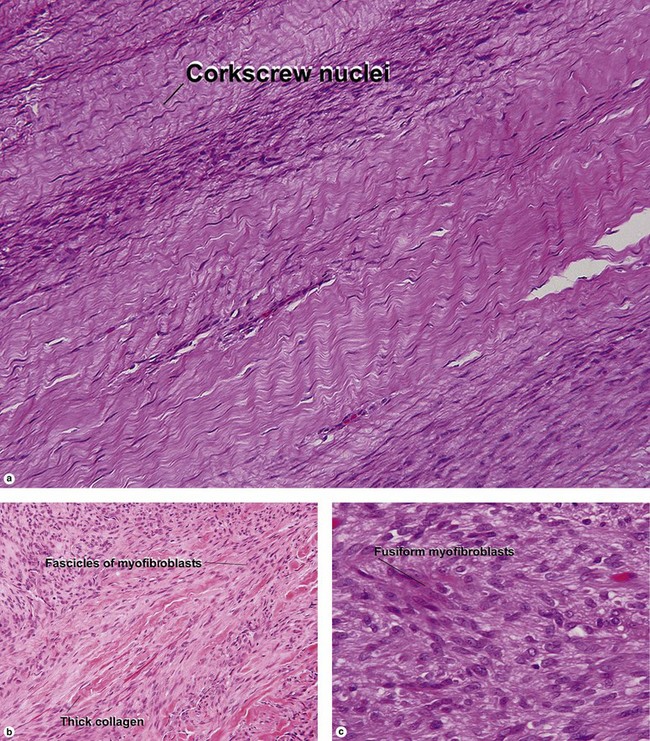
Adult myofibroma
Infantile myofibromatosis
Plastic Surgery Key
Fastest Plastic Surgery & Dermatology Insight Engine