Cutaneous candidiasis and chronic mucocutaneous candidiasis


Cutaneous candidiasis and chronic mucocutaneous candidiasis


Caroline Halverstam and Steven R. Cohen


Evidence Levels:  A Double-blind study  B Clinical trial ≥ 20 subjects  C Clinical trial < 20 subjects  D Series ≥ 5 subjects  E Anecdotal case reports


image


Cutaneous candidiasis


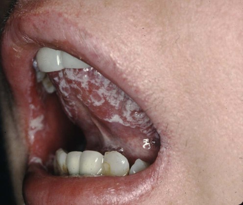
Cutaneous candidiasis is typically caused by Candida albicans, which exists as normal flora of human skin as well as in the gastrointestinal and genitourinary systems. Overgrowth of Candida species is suppressed by normal bacterial flora. Other Candida species occasionally cause mucocutaneous infections, the second most common being Candida tropicalis. Under certain conditions, these Candida species overgrow and become pathogens. Warmth and moisture of the intertriginous skin (axilla, inguinal folds, abdominal creases, inframammary creases), an increased skin pH, and the administration of antibiotics can disrupt the normal bacterial flora, allowing Candida to proliferate. Clinically, candidiasis presents as scaly erythematous patches with satellite papules and pustules. The diagnosis is made either microscopically, with a potassium hydroxide (KOH) preparation revealing spores and pseudohyphae, or by culture.


Management strategy


Topical antifungal agents include, but are not limited to, polyenes, azoles, allylamines, and ciclopirox olamines. Most studies required therapy twice daily for 4 weeks to ensure complete clearance in all patients. Notably, microscopic cure was often present before complete clinical clearance.


Topical corticosteroids are a source of controversy. Although the addition of corticosteroids to local antifungal therapy may reduce local inflammation in acute candidiasis, their use should be limited to 1 or 2 days because of their immunosuppressant properties.


Systemic therapy may be appropriate for cutaneous infections in immunosuppressed patients, in the setting of extensive disease not responding to topical therapy, or in patients non-compliant with topical therapy. Fluconazole 150 mg weekly appears to be as efficacious as fluconazole 50 mg daily or ketoconazole 200 mg daily. As in topical therapy, microscopic cure often precedes complete clinical clearance.


First-line therapies


Second-line therapies


Fluconazole versus ketoconazole in the treatment of dermatophytoses and cutaneous candidiasis.


Stengel F, Robles-Soto M, Galimberti R, Suchil P. Int J Dermatol 1994; 33: 726–9.


Patients were treated with either fluconazole 150 mg once weekly plus daily placebo, or ketoconazole 200 mg once daily plus weekly placebo for 2 to 6 weeks. Candida was present in 21 of the evaluable 150 infection sites. Both treatments were clinically effective, with sites of infection responding equally to treatment with fluconazole and ketoconazole. Mycologic cure rates were seen in 68 of 73 fluconazole-treated patients and in 70 of 77 ketoconazole-treated patients. There were no major differences noted in the eradication rates between the various organisms. However, fluconazole offered the advantage of once-weekly oral administration.

Stay updated, free articles. Join our Telegram channel

Aug 7, 2016 | Posted by in Dermatology | Comments Off on Cutaneous candidiasis and chronic mucocutaneous candidiasis

Full access? Get Clinical Tree

Get Clinical Tree app for offline access