This article discusses an alternative approach to general anesthesia with the use of local anesthesia in minor operating procedure suites when performing in situ decompression of cubital tunnel syndrome for those patients who have mild to moderately severe symptoms and for those who fail to respond to conservative measures. Anterior transposition can easily be performed in the same setting if indicated all with local anesthesia.
Key points
- •
The procedure results in an anterior transposition of the ulnar nerve in the subcutaneous plane while still under local anesthesia.
- •
Patients benefit from walking in and out of a minor procedure suite without any exposure to the risks of general anesthesia.
- •
There is no difference in our patient outcomes between performing cubital tunnel release under local anesthesia in a minor operating setting compared with the results obtained under general anesthesia in a main operating room.
Introduction
Cubital tunnel syndrome is one of the most common nerve entrapment syndromes, second only to carpal tunnel syndrome. It often results in such symptoms as complaints of numbness and tingling in the ulnar half of the hand, and can extend to more severe symptoms, such as wasting of intrinsic muscles. In many cases, treatment of cubital tunnel syndrome requires surgical intervention. Today, the most recognized surgical interventions are in situ decompression, medical epicondylectomy, subcutaneous anterior transposition, and submuscular transposition. Most of these procedures require general anesthesia and an operating room environment.
Cubital tunnel release with local anesthesia
Cubital tunnel syndrome can be treated in many ways including conservative measures, in situ decompression, medial epicondylectomy, direct release with subcutaneous anterior transposition, and submuscular anterior transposition. Cubital tunnel release, with or without anterior transposition, can easily be performed under local anesthesia without the need for intravenous sedation.
Although carpal tunnel decompression is one of the most common operations performed in the world, the cubital tunnel release is much less common because of the belief held by many surgeons that general anesthesia is required. The surgery can be performed with a brachial plexus block or even an intravenous Bier block; however, it is difficult to obtain a complete release proximally, if two blood pressure cuffs are applied to the arm.
The ulnar nerve originates in the brachial plexus and passes through the cubital tunnel. The elbow joint is very dynamic, with a range of approximately 150 degrees. The ligaments over the ulnar nerve stretch and move with elbow motion. Vanderpool and colleagues indicated that there is a significant stretch of the aponeurosis around the elbow with flexion. In full flexion, the cubital tunnel has been shown to be compressed and narrowed by approximately 55%. It is thought that this decreased volume predisposes the ulnar nerve to compression.
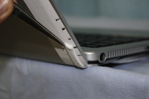
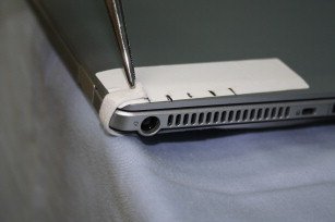
Flexion at the elbow results in the ulnar nerve having to travel a greater distance as opposed to extension of the elbow ( Figs. 1 and 2 ). With elbow flexion, intraneuronal pressure of the ulnar nerve increases significantly. It has also been shown that excursion of the ulnar nerve around the elbow occurs with shoulder and elbow motion. Repetitive motion or dynamic traction and excursion may cause some inflammation around the nerve, which has been validated with histology and imaging studies. Blood flow and axoplasmic flow is affected by compression of the ulnar nerve at the elbow. The ulnar nerve is located superficially at the elbow and mechanical compression may be common when there is very little soft tissue for padding over the nerve. Of course, directly leaning on the elbow and the cubital tunnel can produce a direct mechanical compression on the ulnar nerve. Other sources include soft tissue masses, such as ganglions or lipomas, and direct bony abnormalities, such as cubitus valgus or fractures. Rheumatoid disease, diabetes, and hypothyroidism have all been associated with peripheral neuropathies.
Ulnar nerve anatomy and sites of compression
The ulnar nerve originates in the brachial plexus from C8 and T1 nerve roots. The ulnar nerve containing sensory and motor nerve fibers exits the brachial plexus as a branch of the medial cord and travels from the axillary region to the medial arm.
The first site of compression of the ulnar nerve occurs as it passes posterior to the medial intermuscular septum of the arm. Continuing on its path, the ulnar nerve then passes through the arcade of Struthers, approximately 8 to 10 cm proximal to the medial epicondyle. This arcade lies just anterior to the ulnar nerve and is almost 5 cm in length.
The second site of potential entrapment of the ulnar nerve at the elbow is the medial intermuscular septum. This potential site of compression exists only if there is an anterior transposition of the ulnar nerve or if the ulnar naturally subluxes anterior to the medial epicondyle. As the ulnar nerve approaches the medial epicondyle, it may be entrapped under an anomalous anconeus epitrochlearis muscle, which has been identified as a cause of ulnar compression.
The senior author has performed more than 200 cubital tunnel release procedures and has found that approximately 20% of these patients have had a prominent anconeus epitrochlearis. It is therefore suspected that patients who are symptomatic with ulnar compression at the elbow are more likely to have this anomalous muscle than the general population.
The ulnar nerve then continues toward the elbow and passes underneath the cubital tunnel retinaculum or Osborne ligament. The nerve travels beyond the deep fascia, which is also known as Osborne fascia. The third site of frequent ulnar compression lies deep to Osborne fascia, at the distal aspect of the cubital tunnel.
The ulnar nerve then passes between the ulnar and radial heads of the flexor carpi ulnaris (FCU) muscle. Approximately 5 cm beyond the medial epicondyle, the ulnar nerve penetrates deep to the fascia to lie between the FCU and the flexor digitorum profundus (FDP) muscle bellies. The ulnar nerve provides motor branches to the FCU in this area. The fascial bands between the two heads of the FCU can contribute to the fourth site of compression.
The anterior branch of the medial brachial cutaneous (MBC) nerve and the medial antebrachial cutaneous (MABC) nerve are very important structures in cubital tunnel release surgery. The MBC can sometimes be found just at the proximal edge of the incision, roughly 2 to 5 cm from the medial epicondyle. There is considerable variability in the location of the MABC nerve because it has multiple branches that may pass proximal, over, or distal to the medial epicondyle. These cutaneous branches must be protected when performing cubital tunnel release to avoid iatrogenic injury. The incidence of neuroma formation after cubital tunnel release has been reported to be as high as 30%.
The surgical approaches to cubital tunnel include in situ decompression, which may be performed in an open or endoscopic fashion. The nerve may require anterior transposition to a subcutaneous position or submuscular position. It is difficult to perform a submuscular anterior transposition of the ulnar nerve entirely under local anesthesia.
History and physical examination and investigations
It is important to obtain an accurate history before performing surgery. In cubital tunnel surgery, in particular, it is important to distinguish proximal and distal entrapments, including double crush-type syndrome. Furthermore, it is important to determine if the patient has any further entrapment at the wrist in or around Guyon canal. Document any penetrating trauma, traction-injuries, masses, or history of direct trauma. Other important historical information includes metabolic diseases, such as diabetes, hypothyroidism, and rheumatoid disease. Grading symptom severity is important in terms of evaluating the degree to which the patient has been affected by the disorder.
Importantly, the internal typography of the ulnar nerve explains the relative sparing of the flexi carpi ulnaris and the flexi digitorum profundus because these motor fibers often are found to lie deep within the nerve. The motor branches to the intrinsic muscles are similarly affected in chronic and severe acute compression. The more superficially located sensory fibers are likely to be more susceptible to earlier forms of compression injuries. Tables 1 and 2 respectively describe a nonnumerical grading system of nerve compression at the elbow and a numerical grading system for ulnar nerve compression at the elbow.
| Grade I | Mild symptoms, intermittent paresthsia/hypesthsia, no motor changes |
| Grade II | Persistent symptoms of paresthsia/hypesthsia, varying degrees of mild weakness/atrophy, ulnar innervated muscles |
| Grade III | Persistent sensory symptoms, marked atrophy, or weakness |
| Mild compression | |
| Sensory | Paresthesias come and go, vibratory perception increased |
| Motor | Subjective weakness, clumsiness, or loss of coordination |
| Tests | Elbow flexion or Tinel sign may be positive |
| Moderate compression | |
| Sensory | Paresthesias come and go, vibratory perception decreased of normal |
| Motor | Measurable weakness in pinch or grip strength |
| Tests | Elbow flexion test or Tinel sign are positive, finger crossing might be abnormal |
| Severe compression | |
| Sensory | Paresthesias are persistent, vibratory perception decreased, abnormal two-point discrimination (static ≥6 mm, moving ≥4 mm) |
| Motor | Measurable weakness in pinch and grip muscle atrophy |
| Tests | Positive elbow flexion test or positive Tinel sign may be present, finger crossing may be abnormal |
On physical examination, the ulnar nerve needs to be palpated in flexion and extension for enlargement or subluxation around the medial elbow. Tinel sign is tested over the cubital tunnel and direct manual pressure may be applied over the ulnar nerve for 1 minute to test the ulnar nerve for paresthesias or numbness within the hand. Full elbow flexion with hyperextended wrist may also test the traction on the ulnar nerve.
Motor function, however, is tested through examination of the intrinsic muscles of the hand. There may also be a positive Froment paper sign (ie, flexion of the interphalangeal (IP) joint of the thumb is elicited with a side pinch). Furthermore, a positive Wartenberg sign may be present when there is abnormal abduction of the small finger caused by a weakened third palmar interossei muscle. A distinguishing factor for compression within Guyon canal may be weakness of the extrinsic flexors, such as the FDP.
Contraindications for cubital tunnel release may include a brachial plexus compression caused by a Pancoast tumor or C8-T1 radiculopathy. In the senior author’s experience, these conditions are very uncommon in patients presenting with cubital tunnel syndrome.
Related posts:


Stay updated, free articles. Join our Telegram channel

Full access? Get Clinical Tree


