This article presents the authors’ methods of digital reconstruction using composite tissue transfer. The authors present their approach to achieve restoration of full cosmetic appearance of the reconstructed thumbs or fingers while preserving the function and cosmetic appearance of the donor foot. The reconstructive procedures for each degree of digit defect are discussed in detail, and pitfalls and technical tips are given. This article summarizes the authors’ experience in reconstruction of 646 digits since 1998 and the challenges that they faced in the complex microsurgical reconstruction necessary to pursue the goal of restoring the cosmetic appearance of reconstructed digits and donor feet.
Key points
- •
We present our methods of digital reconstruction using composite tissue transfer.
- •
Our major aims of surgery are to improve the cosmetic appearance of reconstructed hands and donor feet.
- •
We describe several methods using modified or novel designs for composite tissue transfer from the foot to improve cosmetic appearance of the reconstructed digits and to improve both the cosmetic appearance and function of donor feet.
- •
This article provides technical details of the methods we used to move closer to an ideal reconstruction of digits and minimizing donor foot morbidity.
Digital reconstruction: current status and challenges
The loss of a thumb or finger can dramatically affect a person’s work and life. Methods for thumb and finger reconstruction, especially microsurgical techniques, have been extensively studied. However, toe-to-hand transfer is not a perfect procedure in terms of either functionality or cosmetics.
The following are the 2 major challenges:
- 1.
Loss of one or multiple toes from the donor foot: during conventional toe-to-hand transfer, a surgeon harvests the toe according to the length of the damaged thumb/finger. This procedure causes significant damage to the foot and toe that sometimes outweighs the benefit of the reconstructed thumb/finger. Such reconstructive procedures sometimes require the sacrifice of multiple toes. Consequently, unacceptable damage to the foot is created when multiple thumb/finger reconstructions are performed.
- 2.
Poor cosmetics of the reconstructed digits: despite some similarities between thumb/fingers and toes in appearance and function, there are dramatic differences in the lengths of individual phalanges, joints, diameter, and the lengths of a digit from a toe. The reconstructed digits still look like toes, which is especially noticeable when a long digit is reconstructed.
The hand is not only an indispensable tool but also part of a person’s general appearance. Therefore, a digital defect not only impairs a patient’s daily living and work activities but also affects the patient’s self-image and willingness to socialize. Patients often feel embarrassed about showing the digit reconstructed by traditional toe-to-hand transfer techniques.
The focus of our past work on digital reconstruction has been to produce a thumb/finger that closely resembles a normal digit and has good function. At the same time, we have tried to preserve foot/toe function and appearance as much as possible. However, the above-mentioned goals are difficult to achieve through conventional toe-to-hand transfer.
Our methods and goals: ideal cosmetic reconstruction
In 1998, we started to modify the toe-to-hand transfer to produce aesthetic reconstruction of a thumb/finger. To improve the cosmetic appearance of a reconstructed digit, we take different types of grafts from various parts of the body to build a thumb/finger that closely resembles a normal digit.
Here, we will discuss the different surgical techniques that we have used and the results of digital reconstruction in our patients with grade I to VI digital defect.
The goals and unique achievements of our procedures for cosmetic reconstruction of the digit in the hand include:
- 1.
The reconstructed digit closely resembles the corresponding contralateral digit in diameter and length.
- 2.
The lengths of different phalanges, the sizes of nails, and the texture of skin of the reconstructed digit closely resemble the corresponding contralateral digit.
- 3.
The surgical produces cause less damage to the donor foot than previous approaches. Digits can be reconstructed by sacrificing only one toe, or without sacrificing even a single toe.
Clinical patient data
From December 1998 to December 2012, 646 digits (212 thumbs and 434 fingers) ( Table 1 ) were reconstructed with the methods described in this article; 417 digits were on the right hand, and 229 were on the left hand. To preserve the appearance of donor sites, we repaired or reconstructed them as follows:
- 1.
Five toes in the donor foot were preserved in most of these patients.
- 2.
The great toe was used to supply the nail, skin and part of distal phalanx for the reconstruction. The donor soft tissue wound was covered by a pedicle or free flap, and the bone defect was repaired with an iliac bone graft.
- 3.
The second to fourth toes were used to donate the joint and nail. The donor wound was repaired by a pedicle or free flap, and the iliac bone was grafted to fill the bone and joint defect.
| Degree of Defect a | Level of Digital Amputation b | Number of Digits | Number of Patients |
|---|---|---|---|
| I | Distal phalanx | 151 | 136 |
| II | DIP joint (IP joint) | 213 | 197 |
| III | Middle phalanx (proximal phalanx) | 113 | 95 |
| IV | PIP joint (MCP joint) | 93 | 72 |
| V | Proximal phalanx (first metacarpal bone) | 51 | 33 |
| VI | MCP joint (basal joint) | 25 | 19 |
| Total | 646 | 552 |
a Wang CQ, Wang JL, Wang ZT, et al. Thumb and finger reconstruction with the pedal digit transplantation: report of 541 cases. Chin J Repar Reconstr Surg 1997;11:350–2.
b The level of thumb amputation is given in parenthesis if different.
All the 646 reconstructed digits survived completely. The patients were followed up till 2 months after the procedure, at which time the Kirschner (K) wire internal fixation was removed and bone union confirmed. Partial flap necrosis was treated with skin graft and bone graft was performed if non-union of the bone was found. The follow-up 2 months after the procedure varied greatly. Cases involving techniques new to our unit were followed up continuously for 1 to 2 years.
Technical description of our methods
Reconstruction for Grade I Defect
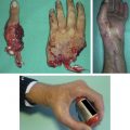
Method 1 – Surgical design: composite tissue transfer from one great toe
According to the defect on the finger ( Figs. 1 and 2 ), a vascularized composite tissue graft including partial nail, dorsal part of the distal phalanx, and a skin flap is taken from the lateral side of the great toe (see Figs. 1 and 2 ). A tongue-shaped flap is left on the medial plantar side of the great toe. Venous return of the harvested flap is through the plantar or dorsal side superficial veins. The lateral plantar digital artery and nerve are used as pedicle for the composite tissue graft.
Harvesting the graft
The dorsal veins are dissected out proximally and divided after reaching appropriate length. They are then dissected out distally toward the lateral side of the tip of the great toe. The plantar digital artery and nerve on the lateral side of the great toe serve as the pedicle for the composite flap. The lateral plantar digital artery and nerve are carefully dissected out to the proximal edge of the flap. The skin is incised on the plantar side of the great toe. The plantar superficial veins are dissected out and divided (see Fig. 1 ; Fig. 2 ). The distal phalanx is exposed, and the periosteum is incised on the lateral side of the nail. A small drill is used to open the cortex on the dorsal and lateral sides of the distal phalanx, and an osteotome is used to separate the lateral from the medial cortex. The partial nail and skin flap are harvested together with the bone based on the same vascular pedicle (see Fig. 2 ). The lateral plantar digital artery and nerve are divided with sufficient lengths.
Resurfacing the donor site wound
Most donor site wounds can be closed directly. However, if the wound is too large, a local advancement flap, in combination with a plantar metatarsal flap, a dorsal metatarsal flap, or a medial-sided flap of the second toe, can be used to resurface the donor site wound.
Preparation of the recipient site
The scar tissue is excised. The digital defect site and nail stump is smoothed with a sharp knife; the bone at the tip of stump is rongeured until the normal bone marrow cavity or cancellous bone is seen. Bilateral digital arteries and nerves are carefully dissected out, and the neuroma at the end of severed nerves is resected. A 1-cm curvilinear incision is made on the dorsal aspect of the middle phalanx of the finger or on the proximal phalanx of the thumb; 1 or 2 dorsal superficial veins are dissected out for anastomosis.
Composite tissue transfer
The vascularized composite tissue from the great toe is transferred to the site of the finger or thumb. The bone is fixed with K-wires, and the nail and nail bed is repaired with 5-0 polydioxanone suture (PDS). Plantar digital artery and nerve from the toe are anastomosed with the digital artery and nerve on the fingers; both dorsal and volar superficial veins are anastomosed (see Fig. 1 ).
Pearls and pitfalls
Owing to the large size of the great toe, only a part of the nail is needed for grade I defect reconstruction. The rest of the nail can be left in situ to preserve the appearance and function of the great toe. Compared with transfer of the second toe to reconstruct the fingertip, a vascularized composite tissue graft from the great toe produces less morbidity on the foot in terms of cosmetic appearance and function. The reconstructed thumb or finger appear and function similar to the missing thumb or finger (see Figs. 1 and 2 ).
The most difficult part of the procedure is dissection of the vein. As no dorsal skin is included in the composite tissue graft, we cannot use the dorsal veins directly for anastomosis. One of the following 3 methods can be used to address this issue: (1) the plantar superficial veins can be used for anastomosis; (2) the dorsal veins on the great toe are identified proximally and dissected distally to the edge of the composite tissue graft, and the distal small branches of the dorsal veins are harvested together with the surrounding soft tissue and do not need to be dissected out individually; (3) a lateral triangular skin paddle can be included in the composite tissue graft, and the veins in the triangular flap can be used for anastomosis with the dorsal veins on the thumb or finger.
Method 2 – Surgical design and procedure: composite tissue from bilateral great toes
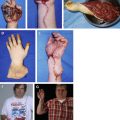
In addition to the method described above, for grade I defect we combine 2 halves of the halluces harvested from both feet to fabricate a thumb or finger and then transfer it to the defect stump ( Figs. 3 and 4 ).
- 1.
The main part of the hallux nail is preserved. The first step is to incise the skin of the finger stump to find the bone, vessels, and nerves. Measure the contralateral finger width of the nail and the circumference of the finger to be reconstructed.
- 2.
Design a composite flap on the lateral sides of both halluces with the length of the flap equal to the finger defect and the width of nail and skin. The width of the harvested toe nail is half of that of the thumb or finger to be reconstructed. The width of harvested flap is decided according to circumference of the reconstruct.
- 3.
Dissect the dorsal skin of the left hallux to find superficial veins, and cut the proximal part to obtain a vein of proper length for the flap. Incise the skin at the proximal line of the fibular side of the flap to show the artery and nerve, which are the artery and nerve for the flap. Incise the nail and the remaining skin as planned, and cut the bone distal to the insertion of the extensor tendon to obtain a part of the phalanx of proper size to match the flap.
- 4.
Harvest the same composite flap from the right foot, and combine the 2 flaps to form a new finger. Fix the fabricated finger on the finger stump with K-wire or thin steel wire. The distal stumps of arteries of the 2 composite flaps are anastomosed to form an artery arch, and the proximal stumps of arteries, veins, and nerves are anastomosed with corresponding arteries, veins, and nerves of the finger stump.
- 5.
Plantar or other flaps near the donor site are used to cover the donor site to decide the foot.
Besides grade I defect, the above method of composite tissues from 2 great toes has also been used for grade II and III defects. From June 2003 to June 2009, 20 fingers that had grade I, II, or III defect were subject to reconstruction surgeries with 1 to 5 years of follow-up. All patients showed normal gait during follow-ups.
Reconstruction of Grade II and III Defects
Surgical design
According to the digital defect and referring to the corresponding digit of the contralateral hand ( Fig. 5 ), a vascularized composite tissue graft (including bone, nail, and skin) is designed on the fibular side of the great toe. As the dorsal skin is included in the composite tissue graft, its dorsal veins are used for anastomosis.
Harvesting the vascularized composite tissue graft from the great toe
- 1.
The dorsal veins are dissected out first and divided proximally at the appropriate length. At the first web space, the lateral plantar digital artery of the great toe, is dissected out. The lateral plantar digital nerve is also carefully dissected out together with the digital artery.
- 2.
The skin flap is elevated, and the dissection is carried out to the bone on the lateral side of the great toe (see Fig. 5 ). The periosteum is incised on the medial side of the nail, and an osteotome is used to open the cortex. The flap is elevated above the extensor tendon up to the distal part of its insertion.
- 3.
An osteotome is then used to cut the dorsal and medial cortex of the distal phalanx. The dorsal lateral part of the bone is separated from the rest of the distal phalanx, which together with a part of the nail and a skin paddle are included in the composite tissue flap. The lateral plantar digital artery and nerve are divided proximally at appropriate lengths.
- 4.
The length of the bone in the composite tissue is measured. An iliac crest bone graft (ICBG) or bone allograft is used if more length is needed (see Fig. 5 ). A vascularized proximal interphalangeal (PIP) joint from the second toe can be used to reconstruct the distal interphalangeal (DIP) joint in the reconstructed finger if necessary.
Donor site wound resurfacing
The secondary wound on the great toe is covered by a pedicled local flap transfer, eg, plantar metatarsal flap, dorsal metatarsal flap, and/or tibial side flap from the second toe (see Fig. 5 ). A free groin flap transfer can also be used.
Transfer to the hand
The vascularized composite tissue is transferred to the finger defect on the recipient hand. A longitudinal K-wire is used for bone fixation. The PIP joint from the second toe can be included with the composite tissue graft to reconstruct the DIP joint of the finger. A conventional iliac bone graft is inserted for additional length requirements. A single longitudinal K-wire is used for bony fixation of the vascularized second toe PIP joint and a conventional iliac bone to the residual phalanx of the recipient digit.
Pearls and pitfalls
- 1.
There is usually enough skin and nail in the vascularized composite tissue graft from the great toe for reconstruction of grade II and III digital defects. However, because of the short length of phalanx from the toe, a conventional iliac bone graft may be needed to restore the length of the reconstructed finger.
- 2.
The secondary wound at the donor site can be covered by a local pedicle skin flap or a free flap such as a groin flap. However, a skin graft is needed to cover the additional wound from the local pedicle skin flap.
Theoretically, the fingers would have better function with the DIP joint motion. However, the DIP joint function in our patients is not satisfactory. Reconstruction of this joint makes the procedure more difficult, and because such a reconstruction for grade II or III finger defect is a challenge, we are uncertain whether it is necessary.
Reconstruction for Grade IV and V Defects
Surgical design
The length, diameter, and location of the joints and the size of the nail on the reconstructed digit are determined by measuring those of the contralateral hand. Based on these measurements, the great toe composite flap and second toe PIP joint transfer were designed on the donor foot. For thumb reconstruction, the diameter of the reconstructed thumb is designed 0.5 to 1 cm larger than the contralateral side, because the reconstructed thumb can become smaller because of atrophy.
Harvesting composite tissue flap from the foot
Preoperative Doppler and computed tomographic (CT) studies reveal the condition of the first dorsal metatarsal artery on the donor foot and the artery on the recipient hand. Designed incisions are made on the donor foot great toe and second toe ( Fig. 6 ). A composite flap including skin, nail, and bone from the big toe and vascularized PIP joint from the second toe are combined to reconstruct the digit. Superficial veins on the dorsal side of metatarsal are exposed and dissected out distally toward the great and second toes. The first dorsal and plantar metatarsal artery, the lateral plantar digital artery and nerve of the great toe, and the medial plantar digital artery and nerve are all carefully dissected out. If the first dorsal metatarsal artery is absent or too small, more length in the plantar metatarsal artery is dissected out as the pedicle. The nail on the great toe is harvested according to the predesigned size. Osteotomy of the toe is performed proximal to the nail root. The lateral half is harvested in the flap. The vascularized PIP joint of the second toe is harvested based on the plantar digital artery with a lateral-sided tongue-shaped flap, with care to protect the dorsal vein and lateral plantar digital artery of the second toe (see Fig. 6 ). Extensor digitorum longus (EDL), flexor digitorum brevis tendons (FDB) to the second toe are harvested according to the size of defect on the recipient site.