Coronal browlift

CHAPTER 24 Coronal browlift


History


The earliest description of coronal brow lifting in the literature was published by Passot in 1919. Since that time, multiple modifications have been made by many talented plastic surgeons including Noel, Lexer, McIndoe, Gonzalez-Ulloa, Vinas, Regnault, Connell, and Ortiz-Monasterio (Paul). Open browlift can be performed using carefully placed incisions within the hair-bearing scalp, anterior hairline, and forehead skin to excise excess skin. Manipulation of the underlying fascia and muscle were well described in the 1950s (Paul). These procedures included surgical fascia reimplantation, myotomies, muscle excision(s), and/or denervation (chemical/surgical). Today, division of the frontal branch of the facial nerve to permanently denervate the frontalis muscle is not recommended since significant brow ptosis has been demonstrated as an unfavorable side effect.


Brow ptosis begins as early as the fourth decade of life. It contributes to sagging of the upper eyelid and most often imparts an aged, sad, and tired appearance to the face. The main etiologic factors in brow ptosis are senescence and gravity. The aging face undergoes a loss of tone from a diminution in the amount of elastic fibers, glycosaminoglycans, and collagen in the skin. Loss of underlying fascial and muscle support occurs, and opposition to the forces of gravity is diminished. Because the lateral brow has fewer attachments to the periosteum and has no underlying frontalis muscle, it usually descends more than the medial brow. Patients typically present with concerns related to a facial appearance that is aged, tired, or sad. Functional sequelae of brow ptosis, such as deficits in the visual field, headaches, or ocular fatigue, are less common.


The open browlift technique allows for direct visualization of sensory nerves and muscles that may otherwise be prone to iatrogenic injury resulting in functional impairment. Proper patient selection can not be overly emphasized.


Anatomy


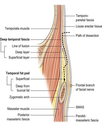
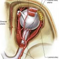
Understanding the temporal and forehead anatomy is critical to successful browlift surgery (Fig. 24.1). The scalp is composed of five layers (Skin, Connective tissue, galea Aponeurotica, Loose areolar connective tissue, and Periosteum). The blood supply to the forehead scalp is from the internal (supratrochlear, supraorbital) and external carotid (superficial temporal) arteries. Hair follicles are located in the subcutaneous layer. Injury to the follicles results in temporary or permanent alopecia. The frontal branch of the facial nerve is located in the superficial temporal fascia and innervates the muscles of the forehead (frontalis, corrugators, depressor supercilii, and procerus). The supratrochlear and supraorbital nerves provides sensation to the central and lateral forehead as well as portions of the anterior scalp.


Stay updated, free articles. Join our Telegram channel

Mar 4, 2016 | Posted by in Aesthetic plastic surgery | Comments Off on Coronal browlift

Full access? Get Clinical Tree

Get Clinical Tree app for offline access