What is the OMT classification system?
The Oberg, Manske, Tonkin classification system is a new system for classification of congenital hand differences. This system was proposed in 2010 and has been recommended to replace the previous IFFSH classification system. The new system reflects deeper clinical and genetic understanding of these conditions. It divides congenital hand differences broadly into three categories: malformations, deformations, and dysplasias.
 What is the most common congenital hand anomaly?
Polydactyly is the most common congenital hand anomaly and it affects approximately 4 to 12 per 1,000 live births.
 What are the three types of polydactyly?
1. Radial polydactyly: Also known as preaxial polydactyly or thumb duplication. Most commonly seen in Caucasian or Asian populations.
2. Central polydactyly: Unusual, comprises only 10% of polydactyly cases. Most often associated with syndactyly (synpolydactyly).
3. Ulnar polydactyly: Also known as postaxial polydactyly. Most commonly seen in African-American or Native American populations.
 What classification system is most commonly used for radial polydactyly? What are the types of radial polydactyly?
The classification system most commonly used is the Wassel classification (Fig. 69-1). Radial polydactyly is classified into six categories based on the level of the polydactyly. The odd numbers indicate polydactyly at the phalanx/metacarpal level, whereas the even numbers indicate polydactyly at the joint level. Type I polydactyly occurs at the distal phalanx, type III at the proximal phalanx, and type V at the metacarpal level. Type II polydactyly occurs at the interphalangeal joint, type IV at the metacarpophalangeal joint level, and type VI at the carpometacarpal (CMC) joint level.
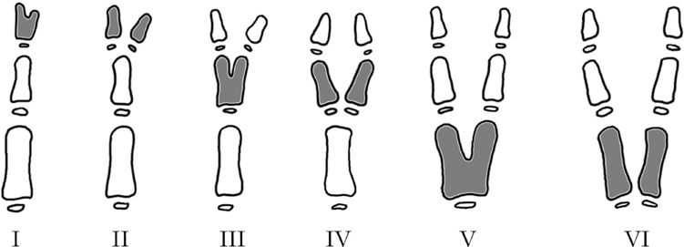
Figure 69-1 Wassel classification for radial polydactyly.
 What is the most common type of radial polydactyly (thumb duplication)?
Type IV; polydactyly occurs at the metacarpophalangeal joint level.
 What are the different types of ulnar polydactyly? Which of these occurs most commonly?
There are three types of ulnar polydactyly. Type I is a soft tissue nubbin with a skin bridge. There is a small neurovascular bundle within this bridge. Type II has skeletal connections with the polydactyly typically occurring at the fifth metacarpophalangeal joint level. Type III is duplication of the entire ray. The most common type is type I.
 Which thumb partner is typically ablated in reconstruction of radial polydactyly? Why?
Usually, the ulnar thumb is preserved and the radial partner is ablated. The main reason to keep the ulnar partner is because it preserves the ulnar collateral ligament of the metacarpophalangeal joint. In practice, the best-looking and most functional thumb is preserved.
 In type IV radial polydactyly, if the ulnar partner is preserved, reattachment of what structure(s) is important to ensure good long-term function?
The radial collateral ligament and the insertion of the thenar intrinsic muscles. Preservation of the ulnar partner implies ablation of the radial partners. When the radial partner is removed, the radial collateral ligament and the insertion of the thenar intrinsic muscles onto the radial side should be preserved. Once the radial partner is discarded, these structures need to be reattached to the ulnar partner to restore balance and centralize the metacarpophalangeal joint.
 What are the long-term unfavorable outcomes after reconstruction of radial polydactyly?
Reconstructed thumbs are usually smaller and stiffer than their normal counterparts. Bulges are possible if the bifid metacarpal or phalangeal heads are not shaved. Deviations are possible if the intrinsic muscle tendons are not rebalanced well.
 What is syndactyly? What are the different types of syndactyly?
Syndactyly occurs when the fingers are fused together—“webbed” digits. It is the second most common congenital hand anomaly. Simple syndactyly indicates fusion of the skin only. Complex syndactyly indicates fusion of the bone and skin. Complete syndactyly extends to the fingertips, whereas incomplete syndactyly terminates more proximally (see Fig. 69-2).
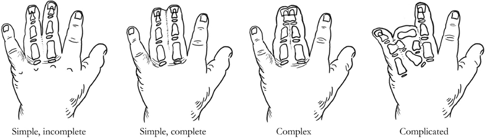
Figure 69-2 Types of syndactyly.
 Which webspace is most commonly involved in syndactyly?
The third webspace.
 What is the embryological timing of digital separation?
The upper limb bud first forms around 4 weeks of gestation. Bones start to appear in the hand around 5 weeks of gestation. Digital rays start to form around 6 weeks of gestational age. At 7 weeks, loose mesenchyme between digital rays starts to break down and notches appear between the rays. By the end of the eighth week, digital separation is complete.
 What is the typical age for syndactyly release? What are the indications for releasing the syndactyly earlier?
There is controversy about the optimal timing for syndactyly release. The typical age for syndactyly release is between 1 and 2 years, and most authors advocate release around 12 to 14 months of age. Release is performed earlier if the syndactyly is causing abnormal deviation or angulation of the involved digits. A typical scenario is syndactyly of the fourth webspace with bony fusion at the distal phalanx. Ring finger growth is restricted distally by a shorter small finger—thus causing ulnar deviation and possible PIP flexion contracture of the ring finger.
 What are the major principles of syndactyly release?
1.
Related posts:
Stay updated, free articles. Join our Telegram channel

Full access? Get Clinical Tree


