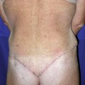
Fig. 32.1
Dehiscence of wound from tight closure
Other contributory causes include smoking, poorly controlled diabetes mellitus, underlying hematoma or seroma, and too much activity by the patient. Many surgeons close the wound tightly with the patient bent tilting the chest and legs toward each other. The surgeon then expects the patient to remain in that position for several weeks even while walking around. Compliance with these instructions is usually poor since it is virtually impossible to maintain this position for any length of time, especially if the patient has a history of back problems.
Treatment for dehiscence should be conservative. It may be possible to debride the wound and do a primary closure. Usually this is not possible because the initial tension was the probable cause of the necrosis and dehiscence. Allowing the wound to slowly slough any necrotic tissue and letting the wound heal by secondary intention is probably the best choice. The necrotic tissue can be debrided at appropriate intervals of time. Usually the scar will contract sufficiently to form a slightly widened scar that can be revised if necessary (if the patient is dissatisfied with the appearance).
32.2.4 Dog-Ears
The closure of the transverse lower abdominal wound is started with a suture in the midline to properly position the new umbilicus. The wound should then be closed starting laterally on each side to give a flattened appearance to the most lateral regions of the wound. If there is excessive fatty tissue in the region where dog-ears usually form, this tissue should be excised prior to the closure. It may be possible to repair the dog-ears at the time of the final closure by liposuctioning the excess fat and then excising the loose skin.
Many times, small dog-ears will resolve within 3 months without treatment. When there are persistent dog-ears for more than 3 months, these can be excised under local anesthesia, usually in an elliptical form. Very large dog-ears and fullness in the area can be treated by liposuction as well as excision.
32.2.5 Edema, Persistent
When edema is persistent, over 3 months, and does not respond to conservative measures such as compression, liposuction with tumescent solution should be considered. Too much liposuction may result in damage to the skin or indentations. Therefore, the liposuction should be conservative. At the same time, there should be a determination as to whether there is persistent edema or just too much residual fat.
32.2.6 Indentation
In raising the abdominal wall flap, fat can be irregularly transected leaving less on some portion of the flap resulting in an indentation. In a modified abdominoplasty, there may be failure to remove fat at the division line between the flap and the remaining abdominal wall to smooth the transition (Fig. 32.2).
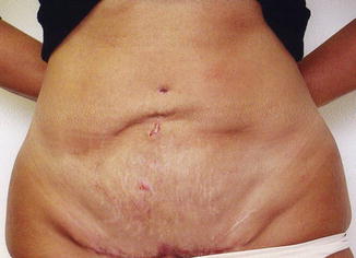
Fig. 32.2
Failure to remove fat at the division line between the flap and the remaining abdominal wall to smooth the transition after miniabdominoplasty
32.2.7 Infection, Sepsis [1]
Wound infection (Fig. 32.3) is a known consequence of any clean surgery, occurring in 1 % of patients in an outpatient or office surgical center and 3 % in a hospital. It is not unusual for slight erythema to occur around the sutures without actual significant infection. If significant wound erythema occurs while the patient is on antibiotics, the dosage may be increased or the antibiotic changed. The wound should respond within 48 h by starting the regression of the erythema. If erythema is persistent or progressing, intravenous antibiotics may be required. This can be given by hospitalization or as an outpatient of the hospital. Infections not responding to antibiotics may require consultation with an infectious disease specialist and may indicate early necrotizing fasciitis or may evolve into that can be fatal. Complete blood count (CBC) should be obtained as well as wound, where possible, and blood cultures. Cultures are best obtained if all the antibiotics are stopped for 48 h.
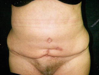
Fig. 32.3
Wound infection resulting in additional scarring
Uncontrolled infection can be life threatening if sepsis occurs. This may be indicated by fever, elevated white count, and lethargy. Prompt treatment with appropriate intravenous antibiotics is essential.
32.2.8 Necrosis [1, 2, 4]
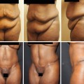
Necrosis is more likely to occur if the patient is a smoker (Fig. 32.4), if concomitant abdominal liposuction is performed, or if there has been prior extensive liposuction to the abdominal wall. Smokers may say they will stop smoking, but there are some who will continue to smoke despite all the admonitions. There is nothing that can be done if the patient does not stop smoking completely. The necrosis will progress to its fullest extent, not only in the lower abdomen but also at times in the periumbilical area and upper abdomen.
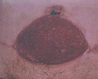
Fig. 32.4
Necrosis of the lower part of the flap and around the umbilicus caused by smoking
Poorly controlled diabetes mellitus, very tight wound closure, underlying hematoma or seroma, and infection may contribute to the cause and extent of the necrosis. If the patient has a prior transverse upper abdominal scar (i.e., cholecystectomy, gastrectomy, splenectomy), the triangle formed by the scar, the midline, and the tightly pulled abdominal skin flap is susceptible to necrosis unless enough space is left between the old scar and the transverse closure line to allow vascularity to the inferomedial triangle. It is helpful to place less tension on the flap closure so that stretching the vessels and thrombosis are not added to the problem. Sometimes, the use of a different type of resection may be necessary to prevent necrosis. This usually means the addition of a vertical midline scar [5].
The best treatment is observation with debridement as needed and allowing the wound to heal by secondary intention. Placing skin grafts in a granulating abdominal wound will shorten the healing time but will not allow the wound to contract enough to decrease the extent of scarring. It is surprising how small the scar can be after complete healing and contraction even if the whole lower abdominal wall has been involved with necrosis. Scar revision is usually necessary for wide or irregular scars.
32.2.9 Necrotizing Fasciitis
Early symptoms are increasing pain, redness and warmth, and general malaise. Swelling then occurs possibly accompanied by a purplish rash. Large violet colored marks appear, and these develop into blisters filled with dark fluid. There may be hypotension and loss of consciousness. Toxic shock syndrome may be associated with necrotizing fasciitis.
Necrotizing fasciitis is a result of infection from Group A Streptococcus or mixed infection, frequently with anaerobic organisms. The infection results in thrombosis of the subcutaneous vessels including vessels entering the fascia and underlying muscles. The tissues become necrotic and require debridement as well as proper antibiotics. Cultures of the tissues may reveal the offending organism(s).
Antibiotics are necessary and cardiac monitoring should be used. Surgery is necessary to remove the necrotic or dead tissues in order to stop the spread of infection. The wound should then be allowed to granulate and can then be skin grafted. Hyperbaric oxygen may help to preserve the healthy tissue.
32.2.10 Need for Further Surgery
There are a variety of reasons for further surgery being necessary following abdominoplasty. These include asymmetry, irregularities, dog-ears, necrosis, inadequate skin resection, significant scar (hypertrophic or keloid), umbilical stenosis, or excessive fat requiring liposuction. If a patient has excessive fat prior to the abdominoplasty and the fat is not liposuctioned at the same procedure, the patient should be informed about the probable future need for liposuction before surgery.
32.2.11 Perforation of Intra-abdominal Organ
It is possible to perforate the bowel when repairing an umbilical hernia or ventral hernia at the same time as performing the abdominoplasty by not opening the hernia sac to observe for attached bowel or placing the sutures superficially only in well-exposed fascia. Closing the midline fascia in a patient with a very loose abdominal wall may require sutures at the lateral edge of the rectus muscle. This is an area consisting only of fascia and peritoneum millimeters in depth where suturing can readily perforate into the abdominal cavity. It might be more appropriate to consider lateral sutures in the external oblique aponeurosis first before central repair so that the central sutures may not need to be placed so far laterally.
Liposuction prior to or after the abdominoplasty may lead to perforation into the abdominal cavity by the cannula. This may injure blood vessels, bowel, or bladder. Perforation of the bowel usually does not become symptomatic until bowel function returns. Then increasing pain occurs from fluid leaking from the bowel into the peritoneal space. Peritonitis is diagnosed by rebound tenderness. Abdominal x-rays may show intra-abdominal air and/or fluid within the peritoneum.
If a perforation is diagnosed, immediate surgical intervention is indicated. Preoperative antibiotics should be started. The abdomen should be carefully explored for possible multiple perforations, and any observed bowel perforations should be closed after thorough irrigation of the abdominal cavity. Early intervention may prevent a severe infectious process.
32.2.12 Recurrent Panniculus
Patients should be forewarned that weight gain after abdominoplasty could result in recurrent fatty abdomen with panniculus that might require another surgical procedure. Pregnancy after abdominoplasty is a risk for causing loose skin, stretching the muscles in the midline, and striae. This may result in the need for repeat abdominoplasty.
32.2.13 Recurrent Protrusion of Abdominal Wall
Some patients have very lax abdominal wall muscles, and there is a tendency for recurrent protrusion after a seemingly adequate fascial repair. This can be improved with repeat closure of the abdominal wall fascia in the midline with the combination of lateral wall (external oblique aponeurosis) tightening. This repair can also be performed for the patient who has recurrent protrusion from loosened or disrupted sutures.
32.2.14 Scarring (Widened, Thickened, Hypertrophic, Keloid) [2]
Wide scars are frequent following abdominoplasty because of the need for a tight closure to get a flat abdomen. When the scar matures and loosens after 6 months, it is possible to resect the scar in order to make it thinner.
Certain individuals are prone to get hypertrophic scars (Fig. 32.5
 Abdominoplasty Combined with Cesarean Section: Discussion of the Evidence
Retrospective Analysis of Never Events in Panniculectomy and Abdominoplasty Patients and Their Financial Implications
Abdominoplasty Combined with Cesarean Section: Discussion of the Evidence
Retrospective Analysis of Never Events in Panniculectomy and Abdominoplasty Patients and Their Financial Implications
 No Drain Abdominoplasty with Progressive Tension Suture: The Logic, Simplicity, and Aesthetics
 Evolution of the High-Superior-Tension Lipoabdominoplasty: The High-Tension Abdominoplasty
 Circumferential Abdominoplasty
 Abdominal Wall Repair Post Hernia in Kidney and Liver Transplantation


Related posts:
Abdominoplasty Combined with Cesarean Section: Discussion of the Evidence
Retrospective Analysis of Never Events in Panniculectomy and Abdominoplasty Patients and Their Financial Implications
No Drain Abdominoplasty with Progressive Tension Suture: The Logic, Simplicity, and Aesthetics
Evolution of the High-Superior-Tension Lipoabdominoplasty: The High-Tension Abdominoplasty
Circumferential Abdominoplasty
Abdominal Wall Repair Post Hernia in Kidney and Liver Transplantation
Stay updated, free articles. Join our Telegram channel
Full access? Get Clinical Tree
