54
Complex Fractures at the Base of the Thumb: Rolando Patterns
John A. Girotto, Shrika Sharma, and Thomas J. Graham
History and Clinical Presentation
The patient presents following an automobile accident with front end impact. At the time of the accident, the airbag deployed. Although the patient does not remember all the details, she thinks she put her hand up when the airbag deployed. She states her thumb is swollen and painful.
Physical Examination
The patient’s thumb is swollen with obvious deformity. The base of the thumb was tender to palpation. Palpation of the carpometacarpal (CMC) joint did not reveal a step-off, but passive movement of the thumb elicited pain. On examination, her left thumb had tenderness at the CMC joint, with a marked prominence of the first metacarpal base. Skin, two-point discrimination, and Allen’s test were intact.
Diagnostic Studies
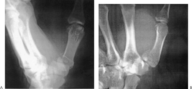
Anteroposterior and lateral radiographs are obtained. They demonstrate the major fragments and the extent of the comminution (Fig. 54–1). Magnetic resonance imaging (MRI) and computed tomography (CT) scans can be helpful in further defining the extent of the fracture if the radiographs are questionable.

Figure 54–1. This patient sustained a closed, comminuted fracture of the thumb metacarpal base as the airbag of an automobile deployed on front-end impact. The anteroposterior (A) and lateral (B) radiographs demonstrate the major fragments and the extent of the comminution.
PEARLS
- Whenever possible, avoid the use of a transarticular wire that is to remain in after the metacarpal fixation.
PITFALLS
- The quality of reduction and the development of later arthritic changes and symptoms have been documented.
Differential Diagnosis
Bennett’s fracture
Metacarpal shaft fracture
Carpal fracture
Rolando’s fracture
Trapezium fractures
Thumb dislocation
Carpometacarpal joint osteoarthritis
Thumb sprain
Diagnosis
Rolando’s Fracture—Intraarticular Fracture of the Base of the Thumb
In 1910, Rolando described a Y-shaped fracture involving the base of the thumb metacarpal that does not result in the diaphyseal displacement characteristic of Bennett’s fracture pattern. Such comminuted intraarticular fractures of the base of the thumb metacarpal are usually in either a Y or a T configuration. Because of the likelihood of posttraumatic arthrosis after these fractures, accurate reduction is important.
In Rolando’s fractures, typically, the radial and ulnar fragments form a T- or Y-shaped pattern. The degree of comminution and the shape of the fragments dictate surgical management of these fractures. If the fragments appear large, exact reconstruction of the articular surface is advocated through an open approach similar to that utilized for Bennett’s fractures. Frequently, however, fractures are found to be more comminuted at the time of operative exploration than was suggested on radiograph.
The first CMC joint is a saddle joint responsible for movement in two axes: flexion/extension and adduction/abduction. Articular embarrassment due to axially loading or hyperextension can generate varying fracture patterns. Injuries to the metacarpophalangeal joint of the thumb are quite common and must be managed appropriately in this important digit. A stable metacarpophalangeal joint is critical for grip and dexterity.
Treatment recommendations for complex thumb base fractures have been varied. They have ranged from a nonoperative approach with aggressive early mobilization to open reduction and rigid internal fixation. As early as 1954, Gedda suggested that exact restoration of the CMC articular surface was necessary for optimal functional results.
When the comminution of the fracture prevents internal reduction and fixation, as in the more complex Rolando’s variants, many authors have advocated external fixation with or without tension band wiring. This technique maintains length and earlier motion. Kontakis et al reported their results with external fixation of Rolando’s fractures. Of 11 patients, only one had unacceptable results secondary to posttraumatic arthritis.
Stay updated, free articles. Join our Telegram channel

Full access? Get Clinical Tree


