95
Common Soft Tissue Tumors/Proliferations
Neural/Neuroendocrine
Neurofibroma
• Skin-colored to pink, soft papulonodule, often on the trunk (see Fig. 50.2).
• Usually solitary in most individuals.
• When multiple, need to distinguish linear form (segmental; mosaic) from a generalized distribution pattern (neurofibromatosis type I) (see Chapter 50).
• Histopathology: wavy, delicate spindle cells with tapered nuclei in a pink stroma.
• Plexiform type has been likened to a ‘bag of worms’ (Fig. 95.1); it is generally on the trunk and proximal extremities, highly associated with neurofibromatosis type I, and prone to malignant degeneration (2–13%).
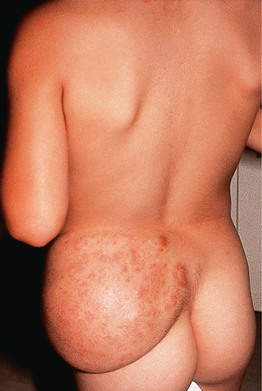
Fig. 95.1 Plexiform neurofibroma in a child with neurofibromatosis. Bag-like mass with overlying patches of hyperpigmentation. Courtesy, Zsolt B. Argenyi, MD.
Schwannoma/Neurilemmoma
Traumatic Neuroma
• Skin-colored papulonodule(s) at a site of prior trauma.
• Often painful or ‘sensitive’ (Fig. 95.3).
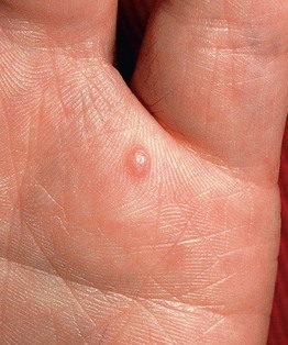
Fig. 95.3 Traumatic neuroma. A painful, firm papule that appeared after a deep puncture injury. Courtesy, Zsolt B. Argenyi, MD.
• Histopathology: haphazardly distributed fascicles of spindle cells with tapered nuclei.
Merkel Cell Carcinoma
• In older adults; solitary, rapidly growing, pink to red to violaceous nodule (Fig. 95.4).
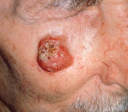
Fig. 95.4 Merkel cell carcinoma (primary cutaneous neuroendocrine carcinoma). Eroded erythematous nodule arising within sun-damaged skin of the cheek. Courtesy, Lorenzo Cerroni, MD.
• Commonly on the head and neck.
Fibrous/Fibrohistiocytic
Angiofibroma (Fibrous Papule)
• Solitary, skin-colored to pink, shiny papule; commonly on the nose (Fig. 95.6).
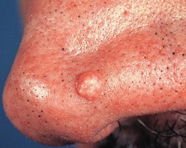
Fig. 95.6 Fibrous papule of the nose. A smooth, dome-shaped, skin-colored papule. Courtesy, Hideko Kamino, MD.
• Histopathology: stellate spindle cells in a hyalinized stroma with dilated vessels.
• DDx: basal cell carcinoma, intradermal melanocytic nevus, adnexal tumors.

