36 Closure Techniques
Summary
Keywords: wound closure single-layer closure trichophytic closure
Key Points
•Preoperative mobility assessment.
•Proper approximation of wound edges and en mass closure of the wound edges.
•Tension-free closure.
•Proper deep placement of the nerve endings.
36.1 Introduction
The history of wound healing dates back to 5,500 to 3,000 BC, the origin of surgery.1 Early suture materials were made of natural materials like silk, linen, cotton, etc. Later, synthetic materials were introduced. The goal of wound closure is to bring the edges of the wound together not only with sufficient strength to prevent dehiscence but also with minimal residual tension and compression of the tissue to promote healing with a cosmetically acceptable scar. In hair restoration surgery, the follicular unit transplantation (FUT) strip scar is of utmost value.
Even today, there is a search for the ideal suture material. The suture materials are broadly classified as naturally occurring and synthetic. These can be monofilament or multifilament (braided), dyed or undyed, coated or uncoated. They can be absorbable, delayed absorbable, or nonabsorbable. All the suture materials have different parameters such as tensile strength, breaking strength, elasticity, capillarity, tissue reaction, ease of handling, knot stability, and absorption rate (Table 36.1).2
Table 36.1 Tensile strength of various suture materials
Suture material | Tensile strength in vivo | Absorption |
Coated Vicryl | 65% at 2 wk | 56–70 d |
40% at 3 wk | ||
Monocryl | 50–60% at 1 wk | 91–119 d |
20–30% at 2 wk | ||
PDS | 70% at 2 wk | >90–210 d |
50% at 3 wk |
The donor wound closure is one of the most important aspects of follicular unit strip surgery (FUSS). A well-healed wound with proper approximation of the wound edges and tension-free closure is likely to result in a very fine scar. There are certain key factors that are prerequisite to the scalp wound closure after strip harvest, that is, mobility of the scalp in the vertical direction, level of strip harvest, and the direction of hair in the upper and lower margins with respect to each other.
The anatomy of the scalp differs in various regions of the head, that is, from the forehead to the superior nuchal lines, there are five layers (skin, subcutaneous tissue, galea aponeurotica, subgaleal loose areolar tissue, and pericranium) and the part below the superior nuchal lines has only three layers (skin, subcutaneous tissue, and deep fascia over the trapezius and sternocleidomastoid muscles).3 This difference can influence the width of the scar tissue after surgical removal. The area above the superior nuchal line allows a wider resection and the movement of neck has little or no effect on wound tension. Moreover, this is also the “safe donor area” in the vast majority of patients. The area above the superior nuchal lines is minimally affected by neck flexion, while the area below the superior nuchal line is impacted during the flexion of neck and therefore will result in a wider scar (Fig. 36.1).
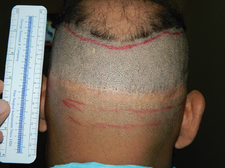
Fig. 36.1 Difference of hair angles in strip surgery.
The ability of the skin to recover from stretch resides in the elastin component. This stretch has little impact on the resultant scar, up until a point at which the limits of collagen fiber extension is reached. At this point, if the force is further pushed, the elastin fibers rupture. These impaired elastin fibers are no longer able to return the collagen to its normal resting state, which results in a permanent consequence called “stretch atrophy.”4
36.2 Single-Layer Closure versus Multilayer Closure
The basic principle of wound closure is to have no tension on the skin edges. The majority of the surgeons use a single-layer closure for a strip harvesting wound (with absorbable or nonabsorbable sutures or staples). There are a few surgeons who prefer a multilayer closure.5 There has been no study to advocate which technique is better in hair transplant surgery. The single-layer closure reduces the surgery time, decreases the exposure of wound surface, and somewhat lowers the cost of the surgery. This can avoid the problems of stitch-related issues, including lower chances of granuloma formation in response to the deep suture material, which in some patients can results in a hypertrophic scar formation. The deeper absorbable sutures get dissolved by the absorption, which may be a nidus for inflammation and thus induce hypertrophic scarring. In addition, some patients who have experienced both single- and double-layer closures state that the deep sutures are more uncomfortable after surgery. The basic aims of multilayer closure are two-fold: to decrease the dead space especially after harvesting a wider strip and to bring the whole flap margin into contact with the other flap margin, thereby reducing the tension on the skin edges.6
The multilayer closure decreases the dead space within the wound, thus decreasing the chances of hematoma formation. It also helps in early removal of skin sutures, which help reduce the chances of cross-hatching. In patients who have scars that are more likely to spread, the deep sutures may help hold the wound together even after the superficial sutures have been removed. The advantages of one-layer running sutures are speed of execution and accommodation of edema during the wound-healing process, but when a donor strip is wide, this technique does not minimize skin tension and may result in the development of a triangle of dead space under the skin suture site.6
Staple closure, as a single-layer closure, is advantageous in that the closure can be completed rapidly, and theoretically causes less damage to the surrounding follicles. However, patients always complain of a significantly greater postoperative discomfort and pain than with sutures as the staples dig into the head when the patient is lying supine.
Subcuticular sutures offer the advantage of avoiding suture marks and may be left in place for several weeks, but the sutures are placed completely in the upper dermis, and because the bulge stem cell region in the follicle is approximately 1 to 1.8 mm below the skin surface, this technique can potentially result in damage to the adjacent hair follicles. To reduce the chance of damage to the surrounding follicles, the surgeon needs to be meticulous to avoid the bulge during suturing. Additionally, absorption of the suture material can potentially cause more reaction during healing and may cause hypertrophy of the resultant scar.
When a multilayer closure is used in other areas of the body, the dermal sutures are used to pull the wound edges together. However, the dermis of scalp is fairly loose, or “cheesy,” and dermal sutures often will cut through the tissue. To avoid this problem, the sutures must be placed deep enough to hold the tissues. The incorporation of galea gives extra strength. The basic aim of multilayer closure is to divert the skin tension to the deeper plane so the secondary effects of tension over the skin margins are avoided.
A two-step suture is mentioned in the literature to reduce the tension of the wound margins. The deeper suture (absorbable) is used subcutaneously fixing the galeal and subgaleal layers. The second suture (nonabsorbable) is used as a modified mattress suture. The key point is to maintain tension within the deep sutures rather than the superficial layer, which may lead to ischemia. A gap of 1 to 2 mm is better than tight skin closure (edge to edge). Minimal tension on wound margins may compensate after edema fluid absorption. Moderate tension may result in telogen effluvium and a wider scar later on. Excessive tension may lead to permanent areas of atrichia, ischemia of the margins resulting in necrosis, or wider scars due to healing by second intension.7
36.3 Absorbable versus Nonabsorbable Sutures
There are various kinds of nonabsorbable suture materials like polypropylene (Prolene), nylon, staples, and absorbable materials like polyglactin (Vicryl), catgut, and poliglecaprone 25 (Dexon).7 The nonabsorbable materials have the least tissue reaction but need to be removed, which can cause patient discomfort, while the absorbable suture materials cannot be used on the skin. These can be used subcutaneously and do not need to be removed. However, these have more tissue reactivity as compared to nonabsorbable sutures. In a study by Bernstein, the superiority of suturing with poliglecaprone 25 as compared to the staples was shown.8 On the other hand, the study by Israr and Stassen reported no difference in healing between staples, silk, polypropylene, and polyglactin for scalp closure.5 The study by Muthuvel et al found staples to be better than sutures in terms of less scar and tissue reaction and potential to conserve hair follicles along the line of the closure; however, the staples were associated with greater patient discomfort.9
36.4 Barbed versus Nonbarbed Sutures
The barbed sutures have a lower rate of absorption and higher suture strength. Barbed sutures can be employed in a simple single-layer closure or a multilayer closure.10 These sutures have been reported to be of value for closure of donor occipital scalp wounds. The nonbarbed (conventional) sutures have various handling issues like securing the knot. The wound tension from suturing may cause tissue ischemia and unfavorable scarring or pressure-induced ischemia and necrosis, leading to wound dehiscence.
36.5 Trichophytic versus Nontrichophytic Closure
The concept of the trichophytic closure was originally used in hair restoration for insetting of the flaps, especially for frontal hairline reconstruction with Juri flaps. The technique can also be used elsewhere. The logic behind the trichophytic closure is to allow the hair growth through the scar, thus eliminating a hairless linear scar and reducing the visibility of the scar. The technique can only benefit those patients in which the mobility of scalp and hair density are adequate. Very lax skin does not benefit from the effect because the scars tend to widen over time. The tricho-closure can be the upper flap, lower flap, or both flaps combined.11,12,13 The choice of the upper margin, lower margin, or double margin remains with the clinical expertise of the surgeon. The tricho-closure involves the removal of the epidermis including one to two rows of hair, approximately 1 mm from the wound margin. The depth of the incision is also about 1 to 2 mm, taking care to stay above the attachment of arrector pili muscles. The removal can be done by sharp scissors or blade. Various techniques have been developed to score the skin, for example, Puig invented a spacer for an additional blade in a multiblade handle; Kim used a “bent” razor blade inserted into a 5-mL syringe as a blade handle, Rose et al mentioned “ledge closure.”14,15,18,19,17,20Frechet described his closure technique in detail, which he used to obtain very fine scars.16 He took smaller strips (<1 cm) above the nuchal ridge in order to have the galea underneath. This intact galea protects the wound from widening. He used no undermining and used a single layer of superficial monofilament suture. The edges of the wound were approximated by the assistant and not by placing tension on the suture. This way the suture closes the wound without “cutting” the tissues or strangulating the follicles. Later, he advocated the use of limited supragaleal undermining. Pathomvanich and Imagawa used towel clamps to both margins of the wound in order to pull them together.13 Application for a few minutes helps in wound closure (Video 36.1).
36.6 Undermining of the Wound Flaps and Use of Bipolar Coagulation Cautery
The use of cautery should be judicial and minimal. The heat produced by cautery can injure the tissues and hair follicles. The debris produced is prone to act like a foreign body and could potentially cause infection. Similarly, the undermining of the flaps causes more tissue response and increases the amount of fibrosis, thus affecting the scalp mobility, which is required in subsequent procedures. It also produces more dead space beneath the flaps, which requires the occlusion by absorbable sutures. In tight scalp or where a wider strip is excised, the undermining is performed to relieve tension where required.16 All these can usually be addressed if the donor area evaluation for mobility is done properly.
36.7 Precautions and Ideal Wound Closure Method
The ideal wound closure method should produce maximal wound eversion and minimal to no follicle damage. It should be tension free and should result in a minimally visible scar. The technique should be easy to perform and should maintain tensile strength throughout the healing process. It should also have precise wound-edge approximation and should not leave stitch marks. The wound should be free of any debris or hair.
36.8 Suturing Technique and Wound Healing
Proper suturing techniques are essential for achieving good cosmetic results and avoiding infection, scarring, and poor wound healing. Techniques that must be mastered include good eversion and precise approximation of skin edges while maintaining uniform tensile strength along the skin edges. The goal of these techniques is to minimize scar formation while avoiding suture marks.
The primary function of a suture is to maintain wound closure and promote wound healing when the integrity of the wound is most vulnerable. The type and amount of suture used, the suturing technique, and the degree of tension on the suture all influence wound healing.
The process of wound healing has characteristically three stages. The initial inflammatory phase (day 0–5) starts as soon as the incision has been created and involves the recruitment of neutrophils and macrophages. The second proliferative phase (day 5–14) is characterized by a decrease in neutrophils and increase in fibroblasts and epidermal cells. The keratinocytes and other epidermal cells migrate to the matrix and proliferate. Adequate moist environment is essential at this stage for re-epithelization. Revascularization also occurs in this phase. The final remodeling phase (day 14 till final healing) is characterized by fibroblasts activity. The myofibroblasts cause wound contraction.
The final wound strength is about 80% of the intact skin almost at the end of 1 year. Only 7% of the final tensile strength is achieved in about 2 weeks increasing up to 20% in 3 weeks. Thus, reducing the tension that a wound experiences during this period will help in better healing in the future months.
In our setting, the sutures (nonabsorbable) are removed after 10 to 12 days. The tensile strength of the wound is around 7 to 10% at this time. If there is tension due to tight closure of the wound placed below the superior nuchal line, it is subjected to the forces, which results in the widening of the scars. If the sutures are removed earlier, chances of wound dehiscence exist, especially in patients undergoing a second session.
The scalp wound can heal by primary intention or secondary intention. The healing by primary intention results in a finer scar, whereas healing by secondary intention results in a larger, atrophic scar. There are various methods used to close the wounds. The one that is obsolete but worth mentioning is “hair tying” and the use of “tissue glue.” Other techniques have been described by Seery,3,6 Brandy, Frechet,16 Pathomvanich and Imagawa,13 Marzola,11 Paul, etc.8
36.9 Authors’ Preferred Technique
36.9.1 Scalp Mobility
An accurate assessment of scalp mobility is a prerequisite in good scalp wound closure technique. Although it is mentioned in previous chapters in the book, the authors put a lot of stress on proper evaluation of scalp mobility as it is a prerequisite to minimal scarring. The wound tension curve becomes vertical and even an additional 1 mm creates a significant problem with closure. The authors use “the vertical scalp mobility scale.”18 The scale is very useful in clinical practice and easy to perform and master. It is found useful especially in the second or subsequent sessions where the width of the strip can be changed accordingly. It also allows the surgeon to have variable width resulting in a tension-free closure and producing a finer scar in the majority of the cases. However, there is never a guarantee of an imperceptible scar because of multiple factors unique to a patient’s healing.
To address the angle of the hair that is changing almost every 1 cm, any strip that is broader than 1 cm is likely to have discrepancies in hair shaft angles in the upper and lower margins (Fig. 36.2). Any discrepancy requires special attention while closing the wound. A wedge suture may be advantageous to reduce the work required to bring the wound edges together when the suture placement angle along the superior edge is increased slightly.19
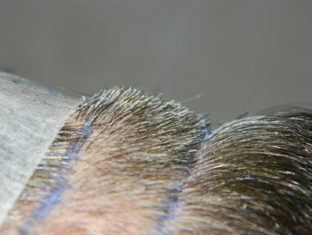
Fig. 36.2 Strip dissection at the subcutaneous level with intact underlying vessels.
Related posts:
 Hair Anatomy and Histology for the Hair Transplant Surgeon
 Plugged in: How to Ensure That Your Practice Thrives (and Survives) in Today’s DigitalWorld
 Transplanting into Areas of Cicatricial Alopecia
 Special Considerations for Postoperative Care in Follicular Unit Excision
 Hairline and Recipient Area Repair of Poor Previous Transplantation
 Ergonomics in Hair Restoration Surgery: FUE Technique
Stay updated, free articles. Join our Telegram channel

Full access? Get Clinical Tree


