Orofacial clefts (OFCs) include a broad range of facial conditions that differ in cause and disease burden. In the published literature, there is substantial ambiguity in both terminology and classification of OFCs. This article discusses the terminology and classification of OFCs and the epidemiology of OFCs. Demographic, environmental, and genetic risk factors for OFCs are described, including suggestions for family counseling. This article enables clinicians to counsel families regarding the occurrence and recurrence of OFCs. Although much of the information is detailed, it is intended to be accessible to all health professionals for use in their clinical practices.
Key points
- •
The following terminology is used when describing orofacial clefts (OFCs): cleft lip alone without cleft palate (CL); cleft lip with or without cleft palate (CL/P), which includes cleft lip only and cleft lip with cleft palate; cleft lip with cleft palate (CLP); posterior cleft palate without cleft lip (CPO); syndromic and nonsyndromic; and familial and nonfamilial (or simplex).
- •
Prevalence is the suggested measure of disease frequency.
- •
The source population, the time period of data collection, the clinical case definition, and the method of case ascertainment are important considerations when comparing prevalence estimates of OFCs.
- •
Both CL/P and CPO may occur in association with other major birth defects. CPO is more commonly syndromic than is CL/P.
- •
The clinical approach includes a history and physical examination for associated morbidity, a thorough gestational history for possible teratogenic factors, and a detailed family history for possible genetic factors.
Historical perspective: terminology and classification
There is considerable ambiguity in the use of terminology when referring to orofacial clefts (OFCs). Many clinicians incorrectly refer to OFCs as deformities, which are said to be the result of disrupted embryologic development. In 1982, an international working group proposed our currently used concepts and terms to describe errors of morphogenesis, which include OFCs. The term malformation should be used for a “morphologic defect of an organ, part of an organ, or larger region of the body resulting from an intrinsically abnormal developmental process.” On the other hand, a disruption is a “morphologic defect of an organ, part of an organ, or a larger region of the body resulting from the extrinsic breakdown of or an interference with, an originally normal developmental process.” In contrast to a malformation, the developmental potential of the involved organ was originally normal and “an extrinsic factor such as an infection, teratogen, or trauma interfered with the development, which thereafter proceeded abnormally.” An example of an OFC caused by a disruption would be one caused by a swallowed amniotic band.
The term deformation (or deformity) should be reserved for “an abnormal form, shape, or position of a part of the body caused by mechanical forces ,” such as plagiocephaly. Although nasal collapse and skeletal asymmetry may be secondary deformities in a child with a repaired cleft lip, the cleft itself is a malformation not a deformity . Finally, the term dysplasia describes “an abnormal organization of cells into tissue s and its morphologic results.” Hence, we have a group of conditions called ectodermal dysplasias , which involve derivatives of the embryonic ectoderm and may have associated OFCs, and skeletal dysplasias , some of which also have OFCs associated with them.
Most OFCs are considered malformations, unless there is clear evidence that it might be a disruption. Even when a cleft is associated with an underlying bone dysplasia or genetic syndrome it is considered a malformation because the process of embryologic tissue growth and fusion was abnormal (because of the underlying syndrome).
The use of the terms isolated and syndromic is another area of potential confusion when describing OFCs. Isolated cleft palate may refer to cleft palate without cleft lip or it may be used to describe a patient who does not have any other malformations or anomalies. In addition, nonfamilial clefts are sometimes called isolated .
The word syndrome means “a pattern of multiple anomalies thought to be pathogenetically related.” A malformation is syndromic if patients have more than one malformation involving more than one developmental field or region of the body. In clinical practice, this usually means birth defects in more than one organ system. For syndromes, pathogenesis is usually unknown, whereas underlying causal factors may be known or unknown.
Some researchers will use the term syndromic in a more restrictive fashion to refer only to patients with syndromes of known or suspected cause (eg, chromosomal syndromes, Mendelian syndromes, eponymic syndromes). The authors suggest calling these syndromes of known cause in order to differentiate them from idiopathic syndromic cases or syndromic cases of unknown cause, which will include OFCs of unknown cause with other major anomalies. A major anomaly is commonly defined as a structural or functional variation from the norm that is of medical, surgical, or cosmetic significance. Both cleft lip with or without cleft palate and posterior cleft palate without cleft lip may occur in association with other major birth defects. Posterior cleft palate without cleft lip is more commonly syndromic than cleft lip with or without cleft palate ( Table 1 ).
| Syndromic (%) | Syndromic with Chromosome Abnormality (%) | Nonsyndromic with Chromosome Abnormality (%) | |
|---|---|---|---|
| CL | 12.1 | 11.3 | 1.8 |
| CL/P a and CLP | 34.6 | 25.0 | 0.6 |
| CPO | 45.9 | 18.1 | 1.6 |
a Some studies did not separate CL from CLP cases. Therefore, this combined category includes some CL cases.
Finally, the difference between sporadic and simplex events should be highlighted. Sporadic refers to a chance event, whereas simplex refers to a single occurrence of a condition in a family. Simplex, or nonfamilial, cases can result from a variety of genetic and nongenetic causes, whereas truly sporadic cases are pure accidents of development. In most cases of simplex OFCs, we simply do not know the cause and, therefore, it is not appropriate to speculate that these are sporadic . As Pagon pointed out, we “need to be very clear when we use the term ‘sporadic’ that it is a chance event with little risk of recurrence and that when we use the term ‘simplex,’ risk of occurrence in relatives remains a possibility.”
Throughout this review, the authors use the terms cleft lip alone without cleft palate (CL), cleft lip with or without cleft palate (CL/P), which includes cleft lip only and cleft lip with cleft palate, cleft lip with cleft palate (CLP), posterior cleft palate without cleft lip (CPO), syndromic and nonsyndromic, and familial and nonfamilial (or simplex) in order to avoid ambiguity.
Classification
Several different classification systems for OFCs have been proposed in the surgical and dental literature. These systems are primarily divided into anatomic systems useful for surgeons and embryology-based systems useful for genetic counseling and research. The disciplines of surgery, genetic counseling, and research require and use different types of OFC data, which has hindered the development of a universally acceptable and useable classification system.
Modern concepts of classification date from the proposal by Kernahan and Stark, which included alveolar ridge clefts with those involving the lip. Based on these concepts a more detailed classification was published. A comprehensive history of OFC classification systems is beyond the scope of this article but has been covered in numerous reviews and classification system proposals.
Animal model data, as well as analysis of recurrence in humans, indicate that, in most cases, CL/P is causally and pathogenetically distinct from CPO. The normal process of secondary palate closure in the human occurs during gestational weeks 6 through 9. Because the lip normally closes by the end of the sixth week and then palate fusion proceeds in a posterior direction over the next several weeks, CPO usually arises from different morphogenetic events than CL/P. Such a view is supported by observations that in families with more than one affected individual, the clefts are usually of the same type. Therefore, traditional empiric recurrence risks given in counseling are risks for CL/P or CPO. There are exceptions to this rule, however, such as the van der Woude and popliteal pterygium syndromes, which are characterized by lower lip pits and either CL/P or CPO. Another example is a Dutch family with 12 affected relatives segregating a causative MSX1 mutation having various combinations of CLP, CPO, and tooth agenesis.
In addition, atypical OFCs include both midline clefts and Tessier oblique clefts, which are relatively rare and are assumed to be both causally heterogeneous and distinct from the more common OFCs.
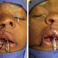
Midline clefts of the lip deserve a special mention. It is extremely important for clinicians to differentiate between the true median cleft lip with a midline, inverted V-shaped notch similar to that normally seen in rabbits, hares, and numerous other animals (apparently the origin of the colloquial term harelip ), and a wide cleft with a flat nose and absent columella caused by premaxillary agenesis. The former, true median or midline cleft, is rare and usually involves only the vermilion border but may extend into the alveolar ridge. This type of true midline notch or cleft is frequently associated with syndromes, such as the oral-facial-digital spectrum and Ellis-van Creveld syndrome. On the other hand, the wide midline cleft of the lip and midline alveolar ridge caused by premaxillary agenesis is a hallmark of the holoprosencephaly spectrum of brain malformations. Holoprosencephaly may be caused by numerous chromosomal abnormalities and single gene mutations. These holoprosencephaly syndromes are usually associated with significant developmental delay and frequently early lethality. Newborns with any of these midline clefts require early consultation by a pediatric geneticist and diagnostic testing to help direct future care and counseling.
Finally, classification needs to take into account milder microform, or forme fruste , examples of OFCs such as small paramedian notches in the upper lip and/or alveolar ridge. A scar on the lip at birth indicates that closure occurred later than usual (ie, a cleft lip healed itself in utero after tissues switched from nonscarring fusion to a scarring type of healing process).
Submucous cleft palate (SMCP) is generally considered to be a microform of CPO, and velopharyngeal insufficiency (VPI) seems to represent the mildest end of the CPO spectrum. This point is illustrated by patients with deletions of chromosome 22q11.2 (DiGeorge syndrome, velocardiofacial syndrome [DGS/VCF]); 69% have a palatal abnormality, more than half of which is VPI, SMCP, or bifid uvula.
Although bifid uvula occurs in the general population as a benign trait, it may also suggest the presence of a submucous cleft. In a study of general pediatric patients with either bifid uvula or an even more subtle small notch in the uvula, most had at least some of the associated anatomic or physiologic pharyngeal abnormalities associated with SMCP. On the other hand, one can have an SMCP without a bifid uvula. The subtlety of these milder microforms ensures that ascertainment for epidemiologic studies will be incomplete and, therefore, these OFCs are usually excluded from epidemiologic research. These microforms do, however, maintain their importance in studies of genetic causation as well as in patient care.
In many epidemiologic studies, CL/P, including both CL and CLP, are studied as a group distinct from CPO because embryology suggests that CLP differs from CL only in severity. Evidence has been presented, however, to suggest that CL may be considered distinct from CLP. In an epidemiologic study of 1.8 million live births in Norway, recurrence risk estimates showed a qualitative difference between the 2 categories of cleft lip (CL and CLP), suggesting that disease severity is not the sole distinguishing factor between the 2 phenotypes. Another study shows a clear separation of risk and transmission patterns between cases with CL and those with CLP.
Measures of occurrence of orofacial clefts
Incidence Versus Prevalence
Incidence and prevalence are traditional epidemiologic measures used to quantify the occurrence of disease in a population. Incidence reflects the transition from health to disease where prevalence reflects both the presence of disease as well as how long a person lives with the disease. Incidence is defined as the number of new cases of disease in a population of individuals at risk for developing the disease in a given time period. This measure can be expressed as either a proportion, with the size of the population at risk in the denominator ( Box 1 ), or as a rate whereby the denominator reflects the time at risk in the population. Prevalence is a static measure, which describes the proportion of cases of disease in the population at a given time point ( Box 2 ). In published studies of birth defects, both incidence and prevalence have been used to describe the frequency of OFCs. Measuring the true incidence of OFCs and other birth defects, however, is virtually impossible because of the difficulties in ascertaining incident cases, which occur early in the first trimester and are often spontaneously aborted, as well as with defining the population at risk (number of conceptions).
Number of new cases of disease/population at risk for disease.
Number of cases of disease/size of the population at a given time point.
Typical OFCs are caused by factors operating during the first 9 weeks of gestation. To measure incidence, one would need to include all embryos during the gestational period when the embryos are at risk for developing an OFC. During this early stage of pregnancy, however, many women are unaware that they are pregnant, and many pregnancy losses go undetected. Thus, it is impossible to quantify the number of conceptions that reach the gestational age when an OFC occurs.
Suggested Measure of Disease Occurrence
Birth prevalence, which in birth defects research is often simply referred to as prevalence , is the suggested measure to quantify the frequency of OFCs at the time of delivery. The authors follow that convention here and use the term prevalence through the remainder of this article. Prevalence is a function of the number of new cases of OFCs as well as survival of the fetus. Prevalence of birth defects, including OFCs, is estimated using the following formula (see Box 2 ). In birth defects research, prevalence is an estimate usually expressed as a ratio measure of the number of cases of OFCs among live births, spontaneous fetal deaths (usually limited to 20 weeks’ gestation or greater), and induced terminations (regardless of gestational age) over the total number of live births in a given time period. The estimate is often expressed per 10,000 live births. For example, a prevalence estimate of 0.001 is typically expressed as 10 cases per 10,000 live births. Because the number of fetal deaths is very small relative to the number of live births, exclusion of fetal deaths from the denominator has little practical influence on the prevalence estimate.
Including both fetal deaths and terminations in the numerator more closely reflects the true prevalence of OFCs at the time of delivery. If one includes only the number of cases among live births, then the true prevalence of OFCs will be underestimated.
Prevalence of Orofacial Clefts
The prevalence of OFCs is often reported as the proportion of children who have CL/P and the proportion of children who have CPO. In a population-based study of approximately 8 million births, the prevalence of CL/P was 9.9 per 10,000 births. These data were collected from 54 birth defect registries across 30 countries between 2000 and 2005. This estimate was consistent with the prevalence of 10.2 per 10,000 births as reported in the United States. The prevalence in Western Europe (12.1 per 10,000 births) was similar to that of the United States, yet the prevalence of CL/P in Japan (20.0 per 10,000 births) was twice that of the United States ( Table 2 ).
| Geographic Region | Cases per 10,000 Births | 95% Confidence Interval |
|---|---|---|
| United States | 10.2 | 9.8, 10.6 |
| Western Europe | 12.1 | 11.1, 13.2 |
| Japan | 20.0 | 18.6, 21.6 |
| Canada | 11.5 | 10.2, 12.9 |
| Australia | 9.7 | 8.5, 11.0 |
Thirty-one percent of the cases of CL/P in the United States were CL. Moreover, in the United States, 75% of children with CL/P during this time period were nonsyndromic (no other malformation or only a minor defect), with 8% of cases being associated with a known syndrome and 17% of cases having multiple malformations. Both of these trends were similar across Canada, Western Europe, Australia, and Japan.
The prevalence of CPO (in the United States between 2004 and 2006) was 6.5 cases per 10,000 live births. The birth prevalence of CPO, excluding chromosomal disorders, differs considerably by geographic region ranging from 1.2 cases per 10,000 births in areas of sub-Saharan Africa to 11.3 cases per 10,000 births in the Oceania region. In Europe and Central Asia, the average birth prevalence of CPO is 6.0 per 10,000 births.
Surveillance Systems
Measures of the prevalence of birth defects are usually estimated from data reported by population-based surveillance systems. Surveillance systems collect, analyze, and report measures of disease occurrence for given populations over time. The method of case ascertainment and the data source for cases may vary by geographic region and depends on the structure of the health care system.
Case finding in surveillance systems may be described as either active or passive. Active surveillance requires intensive effort on the part of the surveillance staff to find cases and confirm diagnoses. Passive surveillance typically involves submission of case reports from given reporting sources directly to the surveillance system and may or may not involve subsequent case confirmation to verify diagnoses. Some surveillance systems use a combination of active and passive surveillance. Common data sources for birth defect case ascertainment include hospitals, physician offices, laboratories, prenatal diagnostic clinics, and administrative data.
Given the potential for variability across surveillance systems, one must consider differences in surveillance methodology when comparing published estimates of the prevalence of OFCs. First one must consider the source population of births that are in the catchment area of the surveillance system. Moreover, the time period in which the data were collected, the clinical case definition, and the method of case ascertainment may influence prevalence estimates and should be considered when interpreting such data.
A source of differential case ascertainment is the underreporting of OFCs that occurs with pregnancy terminations. This underreporting seems to be more common among syndromic OFCs, such as trisomy 13, than nonsyndromic cases. In the United States, the frequency of pregnancy terminations is estimated to be approximately 10%, which limits the impact of these missing cases on the estimate of birth prevalence. In regions where the prevalence of pregnancy terminations is higher, the magnitude of this effect may be greater. Additional factors that influence case ascertainment include errors or lack of specificity in diagnostic coding and failure to report births outside of the catchment area.
Risk factors for orofacial clefts
Sex, Race, and Ethnicity
Prevalence of OFCs differs by sex, race, and maternal age. The prevalence of CL/P among males is approximately twice that of females, whereas the prevalence for CPO is about two-thirds that of females.
The prevalence of all OFCs is less for non-Hispanic blacks compared with non-Hispanic whites. African Americans are 44% less likely to have CL/P compared with non-Hispanic whites. Moreover, the prevalence of CPO was approximately 30% less for African American and Hispanic children when compared with whites. Maternal age younger than 25 years and older than 29 years is associated with an increased risk of OFCs compared with mothers aged 25 to 29 years ( Table 3 ).
| Sex a | Prescription medication; folate antagonists, anticonvulsants, retinoic acid a |
| White non-Hispanic race a | Maternal first-trimester heavy alcohol consumption |
| Maternal age | Prepregnancy diabetes mellitus a |
| Maternal smoking | Maternal obesity (BMI ≥30) a |
Environmental Factors
Most nonsyndromic OFCs are thought to be multifactorial in origin, whereby a combination of genetic and environmental factors is involved. An extensive body of research exists regarding possible causes of OFCs. With the exception of family history, relatively few strong associations are clearly established. The risk factors and the magnitude of their effects are different for CL/P and CPO, supporting the idea that these phenotypes are both causally and pathogenetically distinct entities.
Maternal smoking, particularly at high levels, is consistently associated with an increased risk for OFCs in numerous studies. The risk seems to be stronger for CL/P than for CPO. Furthermore, the association between maternal smoking and OFCs is substantially modified by the presence of specific genetic variants in the mother and fetus, most notably involving transforming growth factor-alpha polymorphisms. Maternal nutritional factors also seem to play a role in the development of OFCs in offspring.
Maternal first-trimester consumption of large amounts of alcohol has been associated with an increased risk for OFCs in several studies, although the results have been somewhat inconsistent. Questions persist about the risks associated with moderate drinking as well as the type of alcohol consumed. In mouse studies, alcohol causes CL/P when administered as early as 8.25 days’ gestation, timing equivalent to early in the fourth week after conception in the human.
Several studies have demonstrated a link between periconceptional intake of multivitamins or folic acid and a reduced risk for both CL/P and for CPO. The evidence for a protective effect of folic acid on OFCs is not as strong as that for neural tube defects. Further research is needed to clarify the optimal dosage, timing, and the potential effect on reducing cleft recurrence. The extent of the protective effect of folic acid on OFCs also seems to be mediated by genetic variants, particularly the methylenetetrahydrofolate reductase (MTHFR) 677T and A1298C polymorphisms.
Several prescription medications have been linked to OFCs when taken during the first trimester. Not surprisingly, folate antagonists and certain other drugs that exhibit antifolate properties are associated with an increased risk for OFCs, both CL/P and CPO. These drugs include anticonvulsants, such as carbamazepine, valproic acid, phenytoin, phenobarbital, and trimethadione. Retinoic acid and certain corticosteroids are also associated with increased OFC risk.
Some maternal diseases and obstetric conditions have been linked to OFCs in the babies of affected women. Prepregnancy diabetes mellitus, but not gestational diabetes, confers an increased risk for several congenital malformations, including CL/P and CPO. Maternal obesity (body mass index ≥30) is associated with an increased risk for both CL/P and CPO in some studies; but the magnitude of the risk is relatively small, with odds ratios around 1.3. The pathogenesis of OFCs associated with diabetes and obesity may be similar because obese patients develop glucose intolerance and increased insulin resistance. Rarely, atypical OFCs may be caused by amniotic bands that deform and disrupt the fetal structures (see Table 3 ).
Several other environmental risk factors have been investigated in relation to OFCs, including chlorinated disinfection byproducts in drinking water; environmental contaminants, such as pesticides, air pollution, and environmental estrogens; and occupational exposures, such as organic solvents. No consistent evidence has been established, however, to indicate these are causally related.
Genetic Factors
OFCs are causally complex phenotypes resulting from the interactions of many different potential factors during development. These factors include programmed and stochastic combinations of genetic and environmental influences, whereby the genetic contribution may be determined by one or more genes. OFCs can occur along with other structural and/or functional birth defects as syndromic clefts or without other major structural or functional developmental anomalies as nonsyndromic clefts . A detailed survey of the genetics of OFCs is beyond the scope of this article but is well covered in several recent reviews.
OFCs, like most other birth defects and common diseases of both childhood and adult life, fit the description of complex disorders. These disorders are conditions whereby a single, causative, completely penetrant gene does not always produce the phenotype. Instead, a combination of effects from more than one gene with or without additional environmental (nongenetic) factors may produce the phenotype. Complex phenotypes are causally heterogeneous, which means that over an extended population, the genetic causes of OFCs include both low frequency (ie, rare), high penetrance, causative alleles and more common, low penetrance, susceptibility alleles interacting with environmental factors. Most of the common disorders of children and adults seem to be complex phenotypes, including most birth defects, intellectual disability, autism, short stature, cancer, diabetes, cardiovascular disease, hypertension, stroke, psychoses, and so forth.
In thinking about malformations, it is helpful to subdivide phenotypes into syndromic and nonsyndromic and then by those of known cause and those of unknown cause (idiopathic). Such an overview of OFCs is shown in Fig. 1 .