Fig. 5.1
(a–e) Sites of predilection of melasma: centrofacial (a), malar (b), mandibular (c), forearm extensor (d), upper chest (e)
Fig. 5.2
(a) Centrofacial melasma, (b) malar melasma, (c) mandibular melasma
Studies done on middle-aged women of Puerto Rican descent (average 44 years old) have shown melasma to be aggravated by sunlight with severe sun damage and epidermal hyperpigmentation on histopathology. Almost all patients had melanophages or melanin in the papillary dermis [8]. Patients with mandibular melasma showed no association with the use of estrogen and progesterone supplements, oral contraceptives, or pregnancy. Most of these patients were of perimenopausal age. Studies suggest that mandibular melasma may be a subset of poikiloderma of Civatte [8].
Melasma of the forearms appears to be a common occurrence and appear in postmenopausal women receiving estrogen supplements. It is observed as sharply demarcated patches with a tinge of erythema. The pigmentation may be macular, speckled, scattered, or confluent. It is postulated that the outer forearm melasma pattern may be due to the higher melanocyte population in this area [9]. The melasma in the mid-upper chest may be observed in combination with facial lesions.
5.3.2 Classification of Melasma According to Severity on Physical Examination
For the purpose of simplicity, melasma presentation in our daily outpatient clinic can be graded as mild (Fig. 5.3a), moderate (Fig. 5.3b), and severe (Fig. 5.3c), using the Melasma Severity Scale (MSS). It rates melasma into four grades: 0 = melasma lesions almost equivalent to the surrounding normal skin or with minimal residual pigmentation; 1 = mild, slightly darker than the surrounding normal skin; 2 = moderate, moderately darker than the surrounding normal skin; and 3 = severe, markedly darker than the surrounding normal skin [10, 11].
Fig. 5.3
Melasma: (a) mild, (b) moderate, (c) severe
Other grading/scoring systems have been in use such as the Melasma Area and Severity Index (MASI) and modified MASI (mMASI). Lately, a Melasma Severity Index (MSI) has been proposed as a new and more practical office-based scoring system [12].
5.3.3 Classification of Melasma Using Diagnostic Tools
5.3.3.1 Light Examination
Under the natural visible light, epidermal, dermal, and mixed pigmentation appears light brown, blue-gray, and deep brown, respectively. Visualization by ordinary visible light is limited and highly subjective. With Wood’s light, the visualization of melasma pigmentation is clearer.
Wood’s light, developed for military use by Dr. Walter Wood in 1903, has found its niche in dermatology practice for cases such as tinea capitis, erythrasma, pseudomonas infections, and porphyria cutanea tarda. For melasma, it has been a practical device to calculate the histological classification of the hyperpigmentation. Under Wood’s light, the contrast in pigmentation is increased if melasma is of the epidermal type, decreased when it is a dermal type, and subtle or slightly perceptible if it is a mixed type of melasma [13]. In patients with very dark skin, contrast is unnoticeable. The abundance of melanin in patients with phototypes V and VI allows absorption of most of the light. The skin appears dark as a whole since only a small amount of light returns back to the eye of the examiner [9]. In recent years, however, studies are being conducted assessing this instrument’s sensitivity, specificity, and accuracy as compared to histopathology, dermoscopy, and confocal microscopy [14].
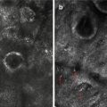
5.3.3.2 Dermoscopy
The use of dermoscopy in melasma has been gaining interest in the past few years. It is a noninvasive technique where the optimal equipment permits a variable magnification from 6× to 400×. Some authors consider the dermoscope as more suitable than Wood’s light in determining the depth of melasma, as the pigment deposition is visualized in an objective manner.
Epidermal melasma shows a brownish hue with regular pigmentary network on dermoscopy. Dermal melasma, on the other hand, presents with a bluish-gray hue with a pigment network that is irregular. Mixed-type melasma shows features compatible to both types. Additional findings such as vascular proliferation have also been appreciated [15, 16]. This is fully discussed in the succeeding chapters.
Related posts:

Stay updated, free articles. Join our Telegram channel

Full access? Get Clinical Tree


