CHAPTER 33 Triceps Augmentation with Silicone Implant
Summary
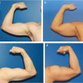
Triceps augmentation with silicone implant is an option for males who seek to have a more muscular physique. While surgery is not without complications, those who seek a more muscular physique may seek harmful methods such as dangerous dieting and anabolic steroids. The evolution of triceps augmentation derived from the works of biceps augmentation. Biceps augmentation was initially developed for upper extremity reconstruction of soft-tissue defects secondary to trauma or oncologic surgery. However, refinements in the surgical technique have facilitated biceps and triceps augmentation to advance from a primarily reconstructive procedure to a powerful tool for cosmetic male biceps and triceps augmentation. Given the rise of males seeking aesthetic surgery, the American Society of Plastic Surgeons showed a 3% increase when comparing 2016 to 2015 and a 28% increase when comparing 2016 to 2000. There will likely be a concomitant increase in males seeking triceps augmentation. In this chapter, we review the pertinent anatomy, preoperative evaluation, operative technique, and postoperative management for triceps augmentation with silicone implantation.
Introduction
The evolution of triceps augmentation derived from the works of biceps augmentation. Biceps augmentation was initially developed for upper extremity reconstruction of soft-tissue defects secondary to trauma or oncologic surgery. In 2006, the use of solid silicone implants for reconstruction of traumatic extremities was reported. 1 In 2010, Chugay and Chugay 2 reported their results of triceps augmentation in 14 patients between 2009 and 2010. Initially, placement of the implant was performed in the submuscular plane but was later transitioned to subfascial placement given improved contour and decreased overall complications. Then, in 2012, Abadesso and Serra 3 reported 32 cases of biceps augmentation using an S-shaped incision in the middle of the arm and placing calf implants into the submuscular plane. In addition, they successfully performed a triceps augmentation in one patient who was pleased with his biceps augmentation. They placed two stacked calf implants beneath the triceps muscle to achieve an augmented triceps.
Increased understanding of male aesthetic anatomy, improved implant design, and refinements in the surgical technique of biceps augmentation have facilitated triceps augmen-tation.
Physical Evaluation
Quality of skin and subcutaneous tissue is examined for laxity and fatty deposits.
Triceps muscle is evaluated at rest and in flexed position while standing; the proximal and distal portions of the long head of the triceps muscle belly are palpated and marked. These markings determine the maximum length of the implant pocket.
Bilateral arm circumference is measured at midportion of arm in a flexed and neutral position to determine degree of triceps hypoplasia.
Asymmetry is noted and pointed out to the patient at the time of examination.
Anatomy
Male upper limb aesthetics are predominantly defined by the shape and development of the triceps and biceps brachii muscles. An anterior and a posterior muscular compartment exist in the upper arm and are separated by the medial and lateral intermuscular septa and humerus. 4 The anterior compartment contains the biceps brachii, brachialis, and coracobrachialis. The posterior compartment contains the triceps brachii muscle. The triceps has three heads: long, lateral, and medial. The long head, the most superficial of the three, arises from the infraglenoid tubercle of the scapula. It extends distally and anterior to the teres minor and posterior to the teres major. The medial head arises distally from the groove of the radial nerve, from the dorsal surface of the humerus, from the medial intermuscular septum; and its distal part also arises from the lateral intermuscular septum. The medial head is mostly covered by the lateral and long heads, and is only visible distally on the humerus. The lateral head arises from the dorsal surface of the humerus, lateral and proximal to the groove of the radial nerve, from the greater tubercle down to the region of the lateral intermuscular septum. The three heads of the triceps then converge into one tendon that begins and inserts into the posterior portion of the olecranon of the ulna.
The deep brachial artery is a large vessel that arises from the lateral and posterior part of the brachial artery, just below the lower border of the teres major, and provides the major blood supply to the triceps. It is relatively superficial throughout its entire course, and care should be taken to preserve it. The venous drainage of the upper extremity is provided by the cephalic vein along with the basilica vein. The ulnar nerve is located in the medial extracompartmental location and runs along the posteromedial aspect of the humerus. Great care should be taken to preserve the ulnar nerve when performing a submuscular dissection.
All three heads of the triceps brachii are innervated by the radial nerve, which arises from the seventh and eighth cranial nerves. In some cases, the long head of the triceps brachii is innervated by a branch of the axillary nerve. The radial nerve is located deep within the posterior compartment adjacent to the posterior aspect of the humerus and medial head of the triceps. The radial nerve and two of its smaller cutaneous branches, the posterior cutaneous nerve of the arm and the dorsal antebrachial cutaneous nerve, are at risk for injury. 5 Great care should be taken to preserve the radial nerve and its branches when performing a submuscular dissection.
The triceps is an extensor muscle of the elbow joint and an antagonist of the biceps and brachialis muscles. The lateral head is used for movements requiring occasional high-intensity force, while the medial fascicle enables more precise, low-force movements. The long head also acts on the shoulder joint and is also involved in retroversion and adduction of the arm.
Patient Selection
The Ideal Candidate
Young patient.
Realistic expectations and goals.
Compliant with instruction.
Lean, very little body fat.
Symmetric upper extremity anatomy.
Good skin quality.
No venous insufficiency.
Implant Goals
A natural result.
Undetectable implant borders.
Matches the other muscle groups.
Upper extremity harmony.
Steps for Triceps Augmentation
In younger patients or more muscular patients with firmer tissues, it is generally recommended to perform biceps and triceps implantation in a staged manner. This allows the largest implant to be placed while avoiding circulation complications postoperatively, particularly the risk of compartment syndrome.
A helpful strategy is to educate the patient about the value of placing multiple implants, which are not “competing for soft-tissue space.” A typical scenario I discussion with my patients is as below:
Stage 1: Biceps, pectoral, medial forearm, medial calf.
Stage 2: Triceps, deltoids, lateral forearm, lateral calf.
Patients tend to appreciate the surgeon’s careful planning, while enjoying the greatest augmentation they can receive, which is appropriate for their soft-tissue envelope.
Implants
Specially designed implants composed of solid silicone, a biologically inert material, is used for triceps augmentation. Given the solid consistency, there is no risk of leakage and systemic dissemination. These implants retain the feel of natural musculature.
Preoperative Markings
Preoperative photographs and videos with the patient flexed and in repose prior to marking are especially helpful to illustrate the asymmetry for the patient. Markings are performed with the patient standing fully upright and the surgeon sitting. In addition to noting the differences between sides and incorporating this into the operative plan, any asymmetry should be pointed out to the patient and clearly documented, as many patients are unaware of these asymmetries. While the implant size and/or shape and pocket position should be adjusted to correct asymmetry as best as possible, preoperative identification is paramount to achieving aesthetic success and for managing patient expectations.preoperative identification is paramount to achieving aesthetic success and for managing patient expectations.
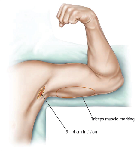
With the arm abducted 90°, the access incision is marked along a natural crease in the axilla. Typically, this incision is 3 to 4 cm long to allow insertion of a folded implant ( Fig. 33.1 ). The triceps contour is then marked out anteriorly and posteriorly with the patient flexed and in repose and additionally with the arm extended. The proximal and distal portions of the long head of the triceps muscle belly are palpated and marked. These markings determine the maximum length of the implant pocket. In addition, the medial head of the triceps is marked out for BodyBanking augmentation. This will give patient the “horseshoe” appearance men desire. Last, areas for concomitant MuscleShadowing are delineated.
Once all areas of interest have been marked and highlighted, the surgeon should stand up and step a few feet back to assess the overall symmetry. Precise markings translate to operative success and have potential to prevent patient dissatisfaction postoperatively. A final set of photographs and videos with the patient flexed and in repose with the markings is obtained.
Related posts:
Stay updated, free articles. Join our Telegram channel

Full access? Get Clinical Tree


