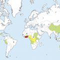
Chagas disease, or American trypanosomiasis, is a parasitic infection caused by the flagellate protozoan Trypanosoma cruzi , an organism that is endemic to Latin America. While Chagas disease is primarily a vector-borne illness, new cases are emerging in non-endemic areas due to globalization of immigration and non-vectorial transmission routes. This article discusses the mode of transmission, evolving epidemiology, pathogenesis, diagnosis, treatment and prevention and control of the disease.
Chagas disease, or American trypanosomiasis, is a parasitic infection caused by the flagellate protozoan Trypanosoma cruzi , an organism that is endemic to Latin America. This disease was first described in 1909 by the Brazilian physician Carlos Chagas, and today the World Health Organization (WHO) estimates that approximately 10 million people are infected.
Although not formally identified until 100 years ago, more recent paleoparasitology studies have revealed the presence of T cruzi DNA in tissue from 9000-year-old pre-Colombian mummies, providing a historical glimpse into an illness that has likely plagued humans for thousands of years and continues to have significant morbidity and mortality and impose far-reaching socioeconomic effects.
Transmission
Chagas disease is primarily a vector-borne illness, with most infections transmitted by blood-sucking reduviid bugs, members of the insect subfamily Triatominae. Triatomines live in the cracks and crevices of poorly constructed mud and thatch homes and emerge at night to procure their blood meal. Most bites are therefore incurred while sleeping and commonly occur on the exposed skin of the face, hence the insects’ common name, kissing bugs. The parasite is transmitted when infected fecal matter from the insect is inoculated into mucosal surfaces or minor breaks in the skin. Within this subfamily, Triatoma infestans , Rhodnius prolixus , and Triatoma dimidiata serve as the 3 most important vectors of human infection. Chagas disease has remained a prolific infectious disease partly because of its large number of mammalian reservoirs, with more than 150 species of domestic, farm, and wild animals (cats, dogs, pigs, rodents, marsupials, armadillos) confirmed as carriers of T cruzi .
Alternate modes of transmission include blood transfusions, solid organ and bone marrow transplants, congenital infection from vertical maternal-fetal transmission, oral infection through food-borne contamination, and rarely, by accidental laboratory exposure.
Historically, transfusion of contaminated blood products has been a common and well-recognized source of T cruzi infections in Latin America. In contrast, only 7 cases of transfusion-associated Chagas disease, all in immunocompromised patients, have been documented in the last 2 decades for the United States and Canada combined. However, the Centers for Disease Control and Prevention (CDC) have reported that nearly 800 cases of Chagas disease have been confirmed in donors at blood donation centers in the United States since 2007, when voluntary screening for this infection was first introduced. Although most of these cases have been concentrated where large populations of Latin American immigrants live, such as California, Texas, Florida, and New York, donor positivity has been documented in 42 states within the contiguous United States.
Similarly, the number of confirmed transplant-associated cases of Chagas disease in the United States also remains small. However, reports of both new infections and reactivation of previous disease have been documented in patients receiving cardiac, renal, and other solid organ and bone marrow transplants in Latin America and represent a growing concern in nonendemic areas.
Another important source of T cruzi infection in both endemic and nonendemic regions is transplacental transmission, with 1 in 20 neonates born to seropositive mothers being diagnosed with congenital Chagas disease. In addition to the concern for congenital infection, Chagas disease also increases the risk of spontaneous abortion and prematurity. Whereas prevention of vertical transmission is not possible, early detection of infection in neonates and prompt treatment greatly reduces the morbidity and mortality.
Although suspected for decades as a potential source of infection, outbreaks of food-borne Chagas disease have been confirmed in recent years in Latin America and are largely linked to contamination of food with fecal matter from triatomine insects. Typically, raw meat or juices derived from sugarcane, guava, or acai berries have been implicated in the oral transmission of T cruzi .
Whereas these modes of transmission represent the minority of cases of Chagas disease, nonvectorial routes are emerging as important sources of new infections, especially in nonendemic regions worldwide.
Epidemiology and Economic Effect
Historically, Chagas disease has affected impoverished rural populations in Mexico and Central and South America, where shoddy construction of domiciles allows the triatomine vectors to thrive. Increasingly, however, Chagas disease is being identified in major cities throughout Latin America, largely a consequence of urbanization and the migration of rural people into metropolitan areas.
The WHO classifies Chagas disease as a neglected tropical disease. This designation implies that the transmission of infection is propagated by poverty, and that the disease often affects vulnerable populations, including indigenous and rural groups, women, children, and the elderly. Furthermore, because poverty favors a greater burden of disease, the large economic effect of this disease in turn contributes to further promoting poverty. Indeed, the economic burden of this disease is significant. In many Latin American countries, the direct and indirect costs, including the cost of health care in dollars and loss of productivity, attributable to Chagas disease ranges from $40 million to in excess of $800 million per nation per annum. Furthermore, as a whole, Latin America experiences economic losses totaling $18 billion annually as a result of the early morbidity and mortality associated with Chagas disease.
An estimated 18 million natives of Mexico and Central and South America have migrated into the United States. Because of this large influx of Latin American immigrants, the CDC estimates that at least 300,000 people currently living within United States are infected with Chagas disease. While most cases of Chagas disease reported in the United States are imported from the endemic regions by immigrants or travelers or transmitted by nonvectorial routes, a handful of vector-transmitted autochthonous infections have been documented in the United States. In fact, triatomine insects are abundant in the Southern United States. A recent study demonstrated that 41% of the insects collected from in and around the homes in Tucson, Arizona were infected with T cruzi , illustrating that the risk of vector-borne transmission of Chagas disease within the United States may be greater than that previously perceived. In addition, several animal reservoirs also exist in many Southern states, evidenced by the identification of T cruzi infection in 20 species of wildlife, particularly raccoons and possums. Of greater concern is the growing number of documented cases of Chagas disease in canines, with numerous cases now reported in dogs from Texas, Tennessee, Louisiana, Oklahoma, Georgia, South Carolina, and Virginia, demonstrating that an active canine transmission cycle for T cruzi exists in the United States. Many features of canine infection parallel the clinical findings in humans, and consequently, Chagas disease confers significant morbidity and mortality on dogs that become infected, requiring veterinarians to increase their recognition of this disease.
Because of the effect of globalization on immigration patterns coupled with the poor recognition of this infection in nonendemic regions and a lack of mandatory screening for all blood and tissue donors, Chagas disease will likely become a growing health threat in the United States. As a result, the current epidemiology of this disease is likely to evolve considerably over the coming decades.
Pathogenesis
The prolific and persistent nature of Chagas disease is likely multifactorial. Without question, the socioeconomic factors, including poverty and substandard housing in rural Latin America, have played a significant role. However, other unique aspects of this parasite’s biology have also contributed, including its large number of mammalian reservoirs, the parasite’s genetic heterogeneity, and a complex cascade of T cruzi -host cell interactions.
Although considerable research effort has focused on these interactions, large gaps in the understanding of T cruzi pathogenesis remain and conflicting theories persist. Most agree that parasite persistence plays an integral role in this disease. Obviously, many complex interactions between the host and parasite dictate the immunopathology of Chagas disease and determine the outcome and success of T cruzi persistence in the human host. For example, molecular mimicry, which promotes autoimmunity, overexpression of parasite peroxiredoxins to counter the host’s oxidative assault, and overexpression of parasitic cysteine proteases such as cruzipain, which trigger a cascade of molecular events resulting in inflammation and tissue damage, are mechanisms that favor parasite persistence. Conversely, a robust T helper cell 1 immune response by the host results in the production of key cytokines, including interferon γ, tumor necrosis factor α, and interleukin 12, along with nitric oxide, which is trypanocidal, and serves to effectively reduce parasite load. Ultimately, the complex web of specific pathways involved in the parasite pathogenesis and human immunopathology of Chagas disease is yet to be fully elucidated, and therefore much of the molecular mystery regarding this infection persists.
Clinical Presentation
Infection with T cruzi has 2 phases: acute and chronic. The acute phase lasts 4 to 8 weeks and is asymptomatic in most infected individuals but may also present as a self-limited febrile illness. If symptomatic, acute Chagas disease manifests as a flulike illness within 1 to 2 weeks of exposure and typically presents with prolonged fever, malaise, anorexia, nausea, vomiting, and diarrhea. When more serious, the patient may display hepatosplenomegaly, lymphadenopathy, and edema. In addition, abnormalities on electrocardiogram (ECG) or cardiomegaly on chest radiograph may be observed, and in rare instances myocarditis, meningoencephalitis, or pneumonitis may develop. Deaths due to acute Chagas disease are seen in less than 10% of symptomatic cases and are attributable to these more severe complications. The risk for fulminant presentations is increased in newborns with congenital infection, children, and immunocompromised individuals.
Of particular note are the local inflammatory reactions at the sites of inoculation, which serve as the earliest and most specific mucocutaneous manifestations of Chagas disease. At cutaneous sites of inoculation, a violaceous, indurated, furuncular nodule with discrete central edema known as a chagoma may develop. If inoculation occurs via the conjunctival mucosae, the patient rapidly develops asymptomatic, unilateral, bipalpebral edema and conjunctivitis with ipsilateral regional lymphadenopathy, a constellation of findings referred to as Romaña sign or ophthalmoganglionar complex. This condition may be complicated by the subsequent development of periorbital cellulitis and metastatic chagomas. Chagomas may persist for several weeks before spontaneously resolving. Finally, a nonspecific and transient morbilliform or urticarial exanthem called schizotripanides may also be observed in the acute phase of the illness.
The manifestations of symptomatic acute phase Chagas disease spontaneously resolve within 1 to 2 months, and the disease transitions into its chronic phase. Chronic Chagas disease is a lifelong infection; however, 60% to 70% of the infected individuals never develop clinically apparent sequalae and are considered to have an indeterminate form of the disease. Conversely, approximately one-third of infected patients experience a latency period of 10 to 30 years, after which they evolve to the determinate form of the chronic phase of Chagas disease, manifesting myriad cardiac, gastrointestinal (GI), and neurologic findings.
Chagasic heart disease is the most common and serious manifestation of chronic T cruzi infection. Conduction system abnormalities, particularly right bundle branch block and left anterior hemiblock, are the earliest signs of cardiac involvement. As the disease progresses, patients develop atrial and ventricular arrhythmias, left ventricular dysfunction, thromboembolic events, dilated cardiomyopathy, and progressive congestive heart failure. Consequently, patients manifest palpitations, syncope, and atypical chest pain. In addition, there is a high risk of sudden death. In fact, cardiac complications are the most common cause of Chagas-related mortality and account for approximately 21,000 deaths annually. The most important prognostic predictors of chagasic heart disease are left ventricular dysfunction, heart failure that meets New York Heart Association functional class III/IV criteria, and presence of nonsustained ventricular tachycardia. Patients with these findings are deemed to be at highest risk for death and should, therefore, be managed aggressively.
GI complications in chronic Chagas disease result from damage to intramural neurons with a direct affect on GI motility. Consequently, patients often experience dysphagia, esophageal reflux, aspiration, abdominal pain, constipation, and weight loss, with the most serious manifestations being megacolon and megaesophagus.
While denervation of parasympathetic nerve fibers is largely responsible for the cardiac and GI manifestations of chronic Chagas disease, primary neurologic complications also occur. The most common findings are altered tendon reflexes and sensorimotor polyneuritis. In addition, stroke secondary to cardioembolic events may represent the first presentation in chronic disease.
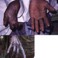
Reactivation of Chagas disease may be observed in immunocompromised individuals, such as those with human immunodeficiency virus infection (HIV) or organ and bone marrow transplants. Although reactivated disease results in easily detectable parasitemia, the clinical presentation differs from that of acute Chagas disease and also differs between those with AIDS and transplant recipients. Specifically, reactivation of Chagas disease in individuals coinfected with HIV is more likely to present with meningoencephalitis and space-occupying brain lesions. By contrast, patients with reactivated disease who undergo transplant are more likely to develop cutaneous lesions of varying morphology including cellulitic plaques, inflammatory indurated nodules and plaques ( Fig. 1 ), ulcers, necrotic eschars, and panniculitis ( Fig. 2 ). In both situations, patients with reactivation are often febrile, acutely ill, and may develop myocarditis.
Diagnosis and Evaluation
Because of the resultant parasitemia during the acute phase of the illness, diagnosis of Chagas disease is made by microscopy, with identification of trymastigotes on peripheral blood smear. However, declining parasitemia in the chronic phase of the disease makes disease detection by microscopy unlikely. Consequently, diagnosis of chronic Chagas disease must be made by confirming the presence of anti– T cruzi IgG antibodies with at least 2 separate serologic tests such as enzyme-linked immunosorbent assay (ELISA), indirect hemagglutination, or indirect immunofluorescence. None of these tests have adequate sensitivity or specificity to be used alone; therefore 2 different methods must be used to sufficiently increase the accuracy of diagnosis. In addition, polymerase chain reaction (PCR)-based methods can provide a definitive diagnosis of Chagas disease during the acute phase of illness, but like microscopy, these methods are limited by the degree of parasitemia and are primarily used for research purposes only.
In cases of suspected transplacental transmission, maternal diagnosis is established by the aforementioned serologic testing methods. To investigate congenital infection, infants born to seropositive mothers should be tested within the first 2 months of life via microscopic examination or PCR testing of cord and/or peripheral blood. If the results of initial testing are negative, follow-up serologic testing is recommended between the ages of 9 and 12 months.
In the United States, 2 detection methods for use as screening tools are approved by the Food and Drug Administration. The first is the ORTHO T cruzi ELISA Test System (Ortho-Clinical Diagnostics Inc, Raritan, NJ, USA) approved in December 2006. Subsequently, a second testing method was approved in April 2010, the ABBOTT PRISM Chagas chemiluminescent immunoassay (Abbott Laboratories, Abbott Park, IL, USA). Both assays are used for qualitative antibody detection in serum and plasma specimens from whole blood, organ, cell, and tissue donors. Neither test is approved for diagnostic purposes.
For seropositive individuals, the initial evaluation should consist of a thorough medical history, review of systems, and physical examination along with a 12-lead resting ECG. For asymptomatic patients with normal findings on ECG, the prognosis is good and yearly reevaluation is advised.
For individuals with abnormalities in ECG or symptomatic disease, subsequent evaluation should be directed by that patient’s specific symptoms or physical examination findings. For example, those with abnormalities in ECG or cardiac complaints should undergo a comprehensive cardiac workup to include 24-hour Holter monitoring, echocardiography, and exercise testing. The findings from these tests dictate the need for additional studies.
For suspected GI involvement, patient should be evaluated by barium swallow and/or with enema. If testing is inconclusive, further studies may be indicated.
In patients with cutaneous lesions, particularly in the setting of Chagas disease reactivation, diagnosis is often possible by skin biopsy. Histologic examination typically reveals a dense lymphohistiocytic infiltrate in the dermis and subcutis, along with numerous intracellular T cruzi amastigotes ( Fig. 3 ), which display prominent kinetoplasts ( Fig. 4 ) and are located within dermal macrophages.
Pathogenesis
The prolific and persistent nature of Chagas disease is likely multifactorial. Without question, the socioeconomic factors, including poverty and substandard housing in rural Latin America, have played a significant role. However, other unique aspects of this parasite’s biology have also contributed, including its large number of mammalian reservoirs, the parasite’s genetic heterogeneity, and a complex cascade of T cruzi -host cell interactions.
Although considerable research effort has focused on these interactions, large gaps in the understanding of T cruzi pathogenesis remain and conflicting theories persist. Most agree that parasite persistence plays an integral role in this disease. Obviously, many complex interactions between the host and parasite dictate the immunopathology of Chagas disease and determine the outcome and success of T cruzi persistence in the human host. For example, molecular mimicry, which promotes autoimmunity, overexpression of parasite peroxiredoxins to counter the host’s oxidative assault, and overexpression of parasitic cysteine proteases such as cruzipain, which trigger a cascade of molecular events resulting in inflammation and tissue damage, are mechanisms that favor parasite persistence. Conversely, a robust T helper cell 1 immune response by the host results in the production of key cytokines, including interferon γ, tumor necrosis factor α, and interleukin 12, along with nitric oxide, which is trypanocidal, and serves to effectively reduce parasite load. Ultimately, the complex web of specific pathways involved in the parasite pathogenesis and human immunopathology of Chagas disease is yet to be fully elucidated, and therefore much of the molecular mystery regarding this infection persists.
Clinical Presentation
Infection with T cruzi has 2 phases: acute and chronic. The acute phase lasts 4 to 8 weeks and is asymptomatic in most infected individuals but may also present as a self-limited febrile illness. If symptomatic, acute Chagas disease manifests as a flulike illness within 1 to 2 weeks of exposure and typically presents with prolonged fever, malaise, anorexia, nausea, vomiting, and diarrhea. When more serious, the patient may display hepatosplenomegaly, lymphadenopathy, and edema. In addition, abnormalities on electrocardiogram (ECG) or cardiomegaly on chest radiograph may be observed, and in rare instances myocarditis, meningoencephalitis, or pneumonitis may develop. Deaths due to acute Chagas disease are seen in less than 10% of symptomatic cases and are attributable to these more severe complications. The risk for fulminant presentations is increased in newborns with congenital infection, children, and immunocompromised individuals.
Of particular note are the local inflammatory reactions at the sites of inoculation, which serve as the earliest and most specific mucocutaneous manifestations of Chagas disease. At cutaneous sites of inoculation, a violaceous, indurated, furuncular nodule with discrete central edema known as a chagoma may develop. If inoculation occurs via the conjunctival mucosae, the patient rapidly develops asymptomatic, unilateral, bipalpebral edema and conjunctivitis with ipsilateral regional lymphadenopathy, a constellation of findings referred to as Romaña sign or ophthalmoganglionar complex. This condition may be complicated by the subsequent development of periorbital cellulitis and metastatic chagomas. Chagomas may persist for several weeks before spontaneously resolving. Finally, a nonspecific and transient morbilliform or urticarial exanthem called schizotripanides may also be observed in the acute phase of the illness.
The manifestations of symptomatic acute phase Chagas disease spontaneously resolve within 1 to 2 months, and the disease transitions into its chronic phase. Chronic Chagas disease is a lifelong infection; however, 60% to 70% of the infected individuals never develop clinically apparent sequalae and are considered to have an indeterminate form of the disease. Conversely, approximately one-third of infected patients experience a latency period of 10 to 30 years, after which they evolve to the determinate form of the chronic phase of Chagas disease, manifesting myriad cardiac, gastrointestinal (GI), and neurologic findings.
Chagasic heart disease is the most common and serious manifestation of chronic T cruzi infection. Conduction system abnormalities, particularly right bundle branch block and left anterior hemiblock, are the earliest signs of cardiac involvement. As the disease progresses, patients develop atrial and ventricular arrhythmias, left ventricular dysfunction, thromboembolic events, dilated cardiomyopathy, and progressive congestive heart failure. Consequently, patients manifest palpitations, syncope, and atypical chest pain. In addition, there is a high risk of sudden death. In fact, cardiac complications are the most common cause of Chagas-related mortality and account for approximately 21,000 deaths annually. The most important prognostic predictors of chagasic heart disease are left ventricular dysfunction, heart failure that meets New York Heart Association functional class III/IV criteria, and presence of nonsustained ventricular tachycardia. Patients with these findings are deemed to be at highest risk for death and should, therefore, be managed aggressively.
GI complications in chronic Chagas disease result from damage to intramural neurons with a direct affect on GI motility. Consequently, patients often experience dysphagia, esophageal reflux, aspiration, abdominal pain, constipation, and weight loss, with the most serious manifestations being megacolon and megaesophagus.
While denervation of parasympathetic nerve fibers is largely responsible for the cardiac and GI manifestations of chronic Chagas disease, primary neurologic complications also occur. The most common findings are altered tendon reflexes and sensorimotor polyneuritis. In addition, stroke secondary to cardioembolic events may represent the first presentation in chronic disease.
Reactivation of Chagas disease may be observed in immunocompromised individuals, such as those with human immunodeficiency virus infection (HIV) or organ and bone marrow transplants. Although reactivated disease results in easily detectable parasitemia, the clinical presentation differs from that of acute Chagas disease and also differs between those with AIDS and transplant recipients. Specifically, reactivation of Chagas disease in individuals coinfected with HIV is more likely to present with meningoencephalitis and space-occupying brain lesions. By contrast, patients with reactivated disease who undergo transplant are more likely to develop cutaneous lesions of varying morphology including cellulitic plaques, inflammatory indurated nodules and plaques ( Fig. 1 ), ulcers, necrotic eschars, and panniculitis ( Fig. 2 ). In both situations, patients with reactivation are often febrile, acutely ill, and may develop myocarditis.
Related posts:
 Buruli Ulcer: Advances in Understanding Mycobacterium ulceransInfection
 Outbreak of Nontuberculous Mycobacterial Disease in the Central Pacific
 Arsenical Keratoses in Bangladesh—Update and Prevention Strategies
 Dermatology in Botswana: The American Academy of Dermatology’s Resident International Grant
 Widespread Use of Toxic Skin Lightening Compounds: Medical and Psychosocial Aspects
Human Immunodeficiency Virus and Leprosy: An Update
Widespread Use of Toxic Skin Lightening Compounds: Medical and Psychosocial Aspects
Human Immunodeficiency Virus and Leprosy: An Update
Stay updated, free articles. Join our Telegram channel

Full access? Get Clinical Tree


