24
Bullous Pemphigoid, Mucous Membrane Pemphigoid, and Epidermolysis Bullosa Acquisita
This chapter, in addition to Chapters 23 and 25, cover the autoimmune bullous diseases. The concepts of direct immunofluorescence (DIF) and indirect immunofluorescence (IIF) are reviewed in Chapter 23 (see Fig. 23.2), as are the recommended sites for performing skin biopsies for DIF (see Fig. 23.1).
Bullous Pemphigoid (BP)
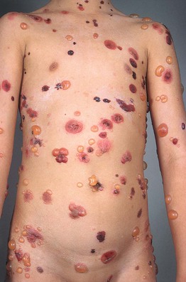
• Both pruritic fixed urticarial plaques and tense bullae are seen (Figs. 24.1 and 24.2); the latter can develop within normal skin or areas of erythematous skin and produce erosions when they rupture; oral lesions are much less common than in pemphigus vulgaris (10–30% of patients).
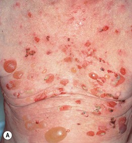
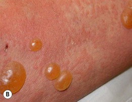
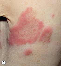
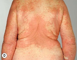
Fig. 24.1 Bullous pemphigoid. A Classic presentation with multiple tense bullae arising on normal and erythematous skin; several of the bullae have ruptured, leaving circular erosions. B Urticarial papules and plaques, along with bullae containing serous fluid. C Firm annular urticarial plaques. D Eczematous presentation with large dermatitic plaques. C, D, Courtesy, Philippe Bernard, MD, and Luca Borradori, MD.
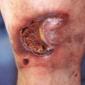
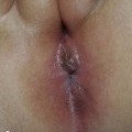
• Pruritus and nonspecific eczematous (see Fig. 24.1D) or papular lesions can precede the more characteristic cutaneous lesions and may be the predominant finding; unusual variants include dyshidrosiform (palms and soles), vegetans (major body folds), and localized (e.g. pretibial in adults; vulvar in children, acral in infants), as well as those that mimic prurigo nodularis and toxic epidermal necrolysis (Fig. 24.3).
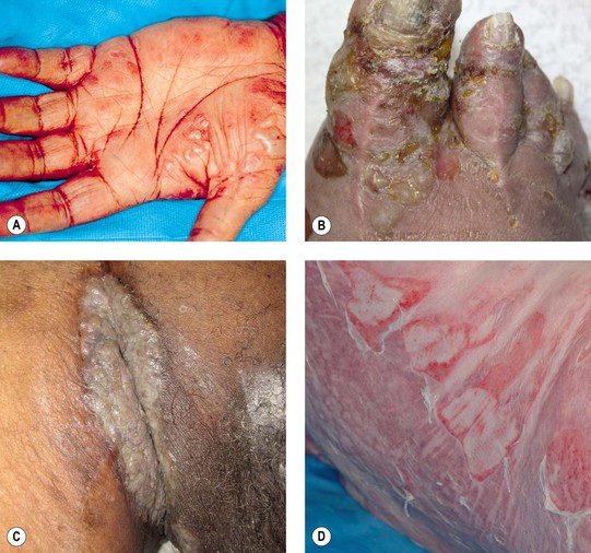
Fig. 24.3 Bullous pemphigoid – unusual clinical variants. Grouped vesicles and bullae on the palms (A) and toes (B) that can resemble pompholyx (dyshidrosiform pemphigoid). C Vegetating plaque in the inguinal crease (pemphigoid vegetans). D Toxic epidermal necrolysis-like lesions with large erosions. A, D, Courtesy, Philippe Bernard, MD, and Luca Borradori, MD.
• Histologically, a subepidermal bulla plus an infiltrate of eosinophils is seen when the lesions are bullous; DIF demonstrates immunodeposits of IgG and/or C3 in a linear array along the basement membrane zone (see Fig. 23.3B); in general, by salt-split skin immunofluorescence studies, the immunodeposits are in the roof (epidermal side) of the blister (Fig. 24.4).
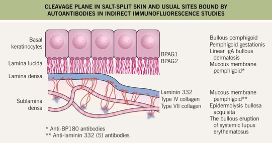
Fig. 24.4
Related posts:
Stay updated, free articles. Join our Telegram channel

Full access? Get Clinical Tree


