Breast reconstruction can be performed safely with local anesthesia. Utilization of the star flap method in conjunction with tattooing successfully provides optimal aesthetic results without the need for an additional donor site. When tissue expander to silicon implant exchange is part of the operative plan, use of triple antibiotic irrigation as well as the Keller Funnel is recommended. Breast augmentation and breast augmentation-mastopexy can also be performed with good results under local anesthetic in a private operating room setting. All other operative conditions, including sterility and sound operative surgical techniques, should be the mainstay of any practice.
Key points
- •
Local anesthetic as an alternative to general or monitored anesthesia care (MAC) has been used for the past 30 years.
- •
Several health risks associated with general anesthesia/MACs are not present with local sedation, providing a safer option for the patient as well as the surgeon.
- •
When attempting to perform surgery under local anesthesia, consider the patient’s desires and tolerance to being awake for the procedure. If anxiety is associated with the procedure or the needle, intravenous sedation is added to the anesthetic plan. A neurologic assessment of the breast mound area is performed, evaluating for light touch and pressure as well as pain.
- •
The star flap and the tattoo method for nipple-areola complex reconstruction, in conjunction with the Keller Funnel sizer to tissue expander exchange to silicon implant performed under local anesthesia, allow a single-site wound and minimal stress, time, and financial burden to the patient, but provide optimal aesthetic results and psychological benefits.
Introduction
As of 2011, more than 93,000 patients were undergoing breast reconstruction. Of those 93,000 patients, two-thirds of the procedures were implant based. More than 60,000 of these patients were postmastectomy breast reconstructions.
Implant-based reconstruction is more frequently performed in 2 stages, with the first tissue expander stage performed immediately after the mastectomies. Results for implant reconstruction have become reliable and have even improved in the setting of radiation therapy in certain instances. Although performing the first stage of reconstruction usually requires general anesthesia, the second stage seems more amenable to performing the procedure under local anesthetic, which can be performed with small incisions with the techniques to be described in this article. Breast augmentation, which is more involved compared with second-stage breast implant reconstruction, has been shown to be performed successfully under local anesthetic.
Reconstruction of the nipple-areola complex (NAC) is most frequently associated with breast cancer and, consequently, mastectomies, and it is also indicated in burn or trauma deformities, complications of reduction mammaplasties, and congenital or developmental disorders. Increase in case numbers over the years has led to many techniques being developed and revised to accommodate the aesthetic objectives of a nipple-areolar reconstruction. The unique texture and color of the NAC makes developing an alternative challenging. Since first being documented by Adams in 1949, the reconstruction of the areola has historically been accomplished via the nonoperative side sharing techniques, grafting from other sites, NAC saving or banking, dermabrasion, and tattooing. Some of these have been used in conjunction with ultraviolet light to facilitate better pigmentation. Reconstruction of the nipple has been achieved through grafting, centrally based flaps, subdermal pedicle flaps, internal nipple prostheses, or autogenous implants. Although skin grafting in conjunction with areolar tattooing can provide an aesthetically pleasing result, it requires a skin graft to be harvested, which in turn produces an additional donor site wound. As an alternative, star flaps in combination with tattooing can provide an equally aesthetically pleasing result without the need for an added site wound.
Local anesthetic as an alternative to general or monitored anesthesia care (MAC) has been used for the past 30 years and provides many benefits to the patient. Several of the health risks associated with general anesthesia/MACs are not present with local anesthetic, providing a safer option for the patient as well as the surgeon. In addition, as an outpatient procedure with no sedation, patients can go home immediately following the procedure, as opposed to general anesthesia/MACs with which the patients must recover from the anesthetic gasses in the postanesthesia care unit (PACU). Furthermore, patients are not required to abstain from the consumption of food or beverages after midnight on the night before their procedure, which alleviates the need for an additional change in the patient’s routine. Patients benefit financially from the procedure being performed under local anesthesia as well. The costs to the patient are significantly less because an anesthesiologist is not necessary for the procedure; recovery in the PACU after stage 1, 2, or both is not necessary; and the only anesthetic is a local medication.
In breast reconstruction, it is therefore feasible to perform tissue expander exchange to permanent breast implant and third stage nipple flap reconstruction under local anesthesia with successful and reliable results. In our practice this is the usual method.
Treatment goals and planned outcomes
Treatment goals for restoring the NAC and exchanging sizers for silicon implants are commonly the same regardless of the surgeon’s technique. Position, size, shape, texture, pigmentation, permanent projection, scar position, and symmetry are essential components for aesthetically pleasing results. The end result must be created in a way that allows patients to readily incorporate the change into their healthy body images. This concentration on the optimization of psychological benefits has been shown to have a positive influence on the overall recovery course of women undergoing postmastectomy breast reconstruction.
Tissue expander exchange to permanent breast implant
Preoperative Planning
The patient is marked in the preoperative holding area with a surgical marker. We use an existing scar to make the incision and generally excise the scar with 1-mm margins in order to provide clean tissue for the subsequent closure. Patients are given 1 dose of prophylactic antibiotics covering gram positives, unless the patient has had a previous infection, in which case we refer to previous cultures to guide our choice of antibiotics. A recent analysis study that searched the literature for antibiotic regimens using 1 dose preoperatively, at 24 hours, and greater than 24 hours showed no significant difference between 24 hours and greater than 24 hours of antibiotic use after surgery. One dose was associated with higher infection rates. However, the literature lacks any randomized trials to answer this question and most plastic surgeons continue to justify their antibiotic protocols based on their training and experience.
Patient Positioning and Procedure
Patients are placed on the operating room table in the supine position with their arms extended out on arm extensions and wrapped with gauze wraps to facilitate sitting the patient up during the procedure to look for symmetry. A chlorhexidine skin preparation is used and, if the procedure requires only a small incision and a simple exchange from a tissue expander to a permanent implants, a small amount of 1% lidocaine with 1:100,000 epinephrine mixture is injected into the incision line and deeper as the surgeon dissects down toward the muscle and capsule/acellular dermal junction. If concomitant revisions of the breast flaps are needed, intravenous sedation and intercostal blocks can be injected, as described by in a recent study. The blocks are injected into the intercostal spaces 3 to 7 with a 1% lidocaine and 0.25% bupivacaine with 1:100,000 equal parts mixture. This mixture is injected at the midaxillary line and the lateral border of the sternum if needed. Because of the added toxicity of both mixtures, we calculate the dose at 4 mg/kg, erring on the lower end for safe dosing.
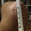
The breast pocket is entered at this junction, which can typically be identified preoperatively by palpation with the patient flexing the pectoralis muscle. The tissue expander is deflated with a #15 blade over suction tubing and removed from the pocket. A minimal incision is used in order for the silicone gel implant to be inserted into the breast pocket. The size of the silicone gel implant is ascertained with silicone gel sizers. In our practice, we have been using the Keller Funnel to introduce the implant into the breast pocket. This funnel allows us to use a smaller incision per implant size. The funnel has standardized markings that represent a specific diameter opening of the funnel ( Fig. 1 ). The funnel end is cut according to the implant size that the surgeon is planning to insert into the breast pocket. Along with changing gloves when handling the implant, and triple antibiotic wash of pocket, implants, and the funnel, a reduction in bacteria load and capsular contracture rates has been reported. The incision is closed using a multilayer closure, and Steri-Strips and sterile gauze dressings are then applied.
Nipple-areola reconstruction
Preoperative Planning and Preparation
Several techniques have been described in nipple reconstruction, including nipple sharing techniques, local flaps, and grafts.
In our practice, we almost always use the modified star flap as described by several investigators. It has offered consistent results and satisfactory outcomes without the need for skin or other tissue grafts and the morbidity that can be associated with those methods. The star flap is also a flap that is easily executed under local anesthetic conditions, circumventing the need for general anesthesia and its associated higher costs and potential morbidity (nausea, vomiting and so forth).
When attempting to perform any operation under local anesthetic, we consider the patient’s desires and tolerance to being awake for the procedure. If there is some anxiety associated with the procedure or the needle, intravenous sedation is added to the anesthetic plan. In addition, a neurologic assessment of the breast mound area is performed, evaluating for light touch and pressure as well as pain. Many patients have decreased or little sensation in this area because of the previous mastectomy and reconstructive procedures.
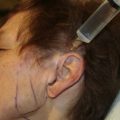
We begin our planning of the modified star flap reconstruction with several key measurements based on specific anatomic points. The suprasternal notch, the midclavicular line, and the inframammary fold are all marked or taken into consideration when placing our nipple. Most of our nipple measurements are within the meridian of the breast and form close to an equilateral triangle in relation to the sternal notch, the nipples on each side, and the lines connecting both nipples on the horizontal. The patient is involved in the final decision of the nipple position by asking her to look into a mirror with us and comment on the placement of the circles we have drawn. If it is a unilateral, the native nipple is used as a reference point. We typically do our mastopexy or reduction symmetry operations in the second stage of an implant breast reconstruction when performing the exchange from tissue expander to permanent implant. The native nipple has therefore settled to a more stable position.
The 3 limbs of the flap are drawn, with the lateral and medial limbs having 2-cm lengths and a 1-cm to 1.5-cm width at their bases. The inferior limb is drawn shorter, to about 1.5 cm, with a width of 2 cm at the base as well ( Fig. 2 ). The inferior limb can sometimes be referred to a superior limb depending on the direction of the blood supply to the flap. As described by Gurunluoglu and colleagues, the horizontal or vertical scar is incorporated into the flap design so that the limb making up the cap of the nipple flap is the one that may cross a preexisting scar. A vertical scar incorporation means that the star flap is designed medially or laterally, and an inferior-based or superior-based flap in relation to a horizontal mastectomy scar.