Disorders With a Characteristic Morphology
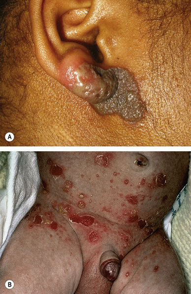
Bullous impetigo (Fig. 14.2) – somewhat tense vesicles/bullae or superficial erosions with collarettes of scale

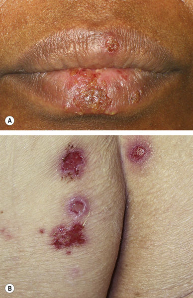
Herpes virus infection (Fig. 14.3) – clustered vesicles and crusts (borders of coalescing lesions may be scalloped), often on an erythematous base; older lesions may be eroded/ulcerated

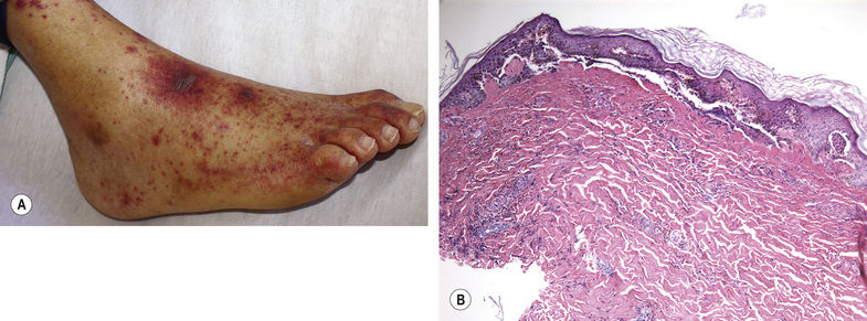
Leukocytoclastic vasculitis (Fig. 14.4) – palpable purpura, sometimes with focal blisters

Fig. 14.4 Leukocytoclastic vasculitis. Purpuric red–purple papules are focally surmounted by bullae. A, Courtesy, William Damsky, MD.

Stay updated, free articles. Join our Telegram channel

Full access? Get Clinical Tree


