“Banana Peel” Scalp, Forehead, and Nape of Neck Flaps
M. ORTICOCHEA
ANATOMY
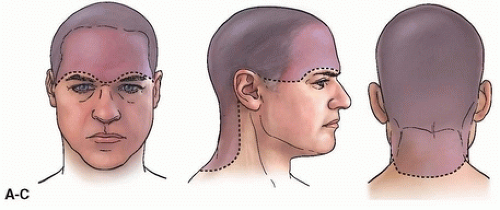
The cutaneous covering of the skull (Fig. 3.1) includes not only the hair-bearing skin, but also the forehead and the nape of the neck. The scalp itself is composed of (1) the hairy skin that covers the skull, (2) the subcutaneous adipose cellular tissue, (3) the underlying epicranial aponeurosis, (4) the subaponeurotic layer of loose connective tissue, and (5) the pericranium (external periosteum).
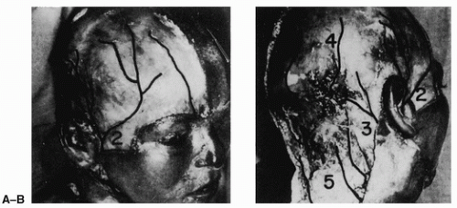
The location of vascular pedicles of the cutaneous covering of the skull is divided into three groups on the basis of which vessels they contain (Fig. 3.2).
Anterior Orbital Pedicle
The anterior orbital pedicle is made up of arteries and veins that reach the skin of the forehead from the orbital cavity and are collateral branches of the ophthalmic vessels. In the middle, they consist of the supraorbital vessels, the most important elements of this group. These vessels reach the forehead via the supraorbital foramen or supraorbital notch of the middle part of the supraorbital border. The notch is easily palpable. This pedicle also includes the supratrochlear vessels that reach the skin on the forehead through the medial part of the supraorbital border. When planning scalp flaps, one should take advantage of the fact that the vessels in this group are located in the central and medial part of the supraorbital border (Fig. 3.2A).
 FIGURE 3.1 Schematic representation of the external limits of the cutaneous covering of the skull. A: Anterior view. B: Lateral view. C: Posterior view. (From Orticochea, ref. 1, with permission.) |
Lateral Pedicle
The lateral pedicle contains the superficial temporal vessels, terminal branches of the external carotid vessels, that reach the scalp after passing in front of the external ear (Fig. 3.2). In front of the superior pole of the ear, they divide into two branches: the frontal branch that proceeds forward, anastomosing with the supraorbital vessels, and the parietal branch that runs upward and backward and ends by anastomosing with the terminal branches of the occipital and posterior auricular arteries.
The superficial temporal vessels are the most important vascular elements to reach the cutaneous covering of the skull because (1) they are the longest vessels of the scalp and thus irrigate a large area of it, and (2) their easy mobilization allows them to be included in a variety of flaps (see Fig. 3.6).
The posterior auricular vessels, collateral branches of the external carotid vessels, reach the scalp just behind the external ear (Fig. 3.2B). These vessels pass the mastoid process of the temporal bone, branching through the scalp in back and above the external ear. They anastomose with the parietal branch of the superficial temporal vessels and with the occipital vessels. The posterior auricular pedicle is less important than the superficial temporal one, because it is of smaller caliber and has shorter branches that are mobilized with difficulty during surgery, since they are firmly attached in the mastoid auricular region.
 FIGURE 3.2 A,B: Schematic representation of the circulation of the cutaneous coating of the skull: (1) supraorbital and supratrochlear vessels; (2) superficial temporal vessels; (3) posterior auricular vessels; (4) lateral and medial branches of occipital vessels; (5) multiple perforating vessels, which are collateral branches of the occipital, vertebral, and superficial cervical arteries. (From Orticochea, ref. 1, with permission.)
Stay updated, free articles. Join our Telegram channel
Full access? Get Clinical Tree
 Get Clinical Tree app for offline access

|


