24
Arterial Aneurysms
Kevin D. Plancher
History and Clinical Presentation
A 38-year-old left hand dominant woman is seeking treatment for a tender mass in the palm of her hand. The patient had fallen with a glass bottle in her hand 2 weeks earlier and had sustained a puncture wound to the ulnar aspect of her right palm. At that time active arterial bleeding persisted despite direct pressure on the wound with a compressive dressing. She was originally treated in the emergency department with irrigation and exploration with cauterization of a “superficial” vessel and wound closure.
Physical Examination
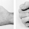
Removal of her sutures of the skin resulted in pulsatile bleeding from the wound. The bleeding continued despite direct manual compression on the wound (Fig. 24–1). Manual compression of the ulnar and radial arteries individually at the wrist crease did not stop the bleeding. Bleeding only ceased with manual compression of both arteries. The Allen’s test was performed and found the ulnar artery not patent. The color of the hand when performing the Allen’s test, to the ulnar side of the wrist, showed lack of perfusion to the radial side of the wrist (Fig. 24–2).
PEARLS
- Use of a tourniquet and appropriate anesthesia for adequate visualization and microscopic dissection of the artery
- Meticulous microscopic dissection with tension-free anastomosis
- Postoperative supervised therapy and appropriate incision care
PITFALLS
- Vein graft selection too small
- Inadequate graft length or anastomosis sewn under tension
- Failure to maintain proper graft orientation
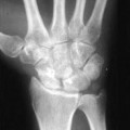
Diagnostic Studies
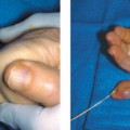
Radiographs of the patient’s hand were obtained. Doppler flow assessment, pulse volume recordings, and angiography were also obtained. Angiography provided definitive information regarding the location and extent of arterial injury (Fig. 24–3).
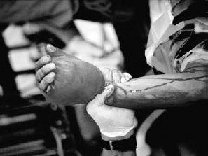
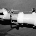
Figure 24–1. Manual compression of the wrist with a saturated dressing in the patient with persistent bleeding.
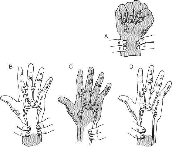
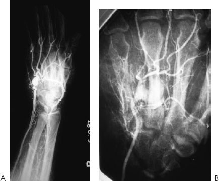
Figure 24–3. (A) Arteriography of the forearm and digital subtraction angiography of the palm. (B) Close-up view of the palm.
Differential Diagnosis
Ganglions
Nerve tumors
Other soft tissue masses
Raynaud’s disease
Pseudoanerysm of the palm
Diagnosis
Pseudoaneurysm of the Palm
Aneurysms are classified into two groups: traumatic and nontraumatic. Traumatic aneurysms can be subgrouped into true and false aneurysms. True aneurysms occur after blunt trauma to the upper extremity. These arise from occupational hazards, thoracic outlet syndrome, or a malignancy. Damage occurs to the arterial wall media to cause vessel dilation.
Related posts:
Stay updated, free articles. Join our Telegram channel

Full access? Get Clinical Tree


