Arsenic is considered a Class I human carcinogen by the International Agency for Research on Cancer because of its increased risk for skin cancer, as well as internal cancers, such as lung and bladder cancer. Arsenic contamination of drinking water in Bangladesh has been called the “largest mass poisoning of a population in history.” This inorganic arsenic contamination is of natural origin, with arsenic thought to be released to the groundwater from the surrounding sediment. Arsenicosis and its risk factors and prevention and management are discussed in this article.
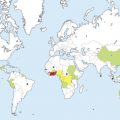
Arsenic contamination of drinking water in Bangladesh has been called the “largest mass poisoning of a population in history,” a consequence of the widespread installation and use of tube wells intended to provide a safer alternative to surface water sources that could easily be contaminated by microorganisms. It has been estimated that by the 1990s, 95% of the rural population of Bangladesh was drinking water from tube wells.
The current World Health Organization (WHO) guideline designates 0.01 mg/L as the maximum permissible amount of arsenic in drinking water. The National Arsenic Mitigation Information Center reported in 2008 that of 4.8 million tube wells evaluated by field testing kits in Bangladesh, almost 30% had arsenic levels exceeding 0.05 mg/L. This inorganic arsenic contamination is of natural origin, with arsenic thought to be released to the groundwater from the surrounding sediment. A recent survey by Chakraborti and colleagues estimated that in Bangladesh, 36 million people are at risk of drinking arsenic-contaminated water at arsenic levels more than 0.01 mg/L, with 22 million at risk of drinking water with arsenic levels more than 0.05 mg/L. Previous estimates put the population at risk for arsenic exposure at levels beyond the WHO guideline to be as much as 35 million to 57 million.
Chronic arsenicosis is defined by the WHO as a “chronic health condition arising from prolonged ingestion of arsenic above a safe dose for at least 6 months, usually manifested by characteristic skin lesions of melanosis and keratosis, occurring alone or in combination, with or without involvement of internal organs.” The latency for the appearance of clinical signs and symptoms of chronic exposure can range from 6 to 10 months or even 20 years or more. Extracutaneous manifestations include neurologic changes, hypertension and cardiovascular disease, pulmonary disease, peripheral vascular disease, diabetes, adverse pregnancy outcomes, and internal malignancy. Arsenic is considered a Class I human carcinogen by the International Agency for Research on Cancer because of its increased risk for skin cancer, as well as internal cancers, such as lung and bladder cancer. Proposed mechanisms for arsenic carcinogenesis include chromosome abnormalities, oxidative stress, altered growth factors, cell proliferation, promotion and/or progression in carcinogenesis, altered DNA repair, p53 gene suppression, altered DNA methylation patterns, and gene amplification. At very high concentrations of exposure, systemic symptoms, most commonly gastrointestinal symptoms as well as peripheral neuropathy, may precede skin lesions. Recently evaluated data from the Health Effects of Arsenic Longitudinal Study (HEALS) show that the risk of all-cause mortality and chronic disease mortality increased with increasing arsenic exposure, particularly long-term exposure.
Cutaneous disease
The first case of arsenicosis was identified in a patient in Bangladesh as early as 1984, and a recent study found patients with skin lesions in 31 of 33 surveyed districts in Bangladesh. Arsenic-related skin lesions are considered the earliest manifestation of nonmalignant disease related to chronic arsenic exposure. The risk for nonmalignant and malignant skin disease seems to increase with increasing dose. One study noted that subjects exposed to an arsenic concentration of 0.05 mg/L had a 59% higher risk of skin lesions compared with unexposed subjects. Skin changes can be found with chronic exposure to nonlethal doses of arsenic, ranging from 0.005 to 0.09 mg/kg/d.
Nonmalignant and Premalignant Disease
Of the skin findings occurring in arsenicosis, pigmentary changes are considered to be the earliest dermatologic findings. These alterations can be diffuse or patchy and are often localized to the palms and soles. Characteristic patterns include a freckled raindrop pattern, leucomelanosis (guttate hypopigmentation in a background of hyperpigmentation), and mucosal pigmentation. Conjunctival congestion and nonpitting edema have also been demonstrated.
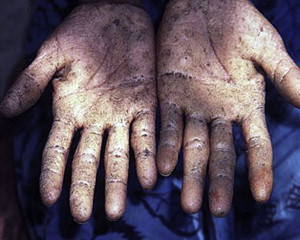
The presence of arsenical keratoses has been suggested to be a sensitive marker for early detection of arsenicosis and is the most common manifestation preceding the development of arsenic-related skin cancer. Typical lesions are hyperkeratotic, punctate, firm papules measuring 2 to 10 mm in diameter and are often located at sites subject to friction or trauma ( Fig. 1 ). Common locations are the palms and soles, although they may also be present on the dorsal surface of the extremities ; other locations include the trunk, genitalia, and eyelids. Arsenical keratoses also may present as scaly, erythematous, or hyperpigmented plaques. Thickened leathery plaques may have associated hyperhidrosis. The extent of disease is graded on a scale of mild, moderate, and severe. In mild disease, lesions are usually less than 2 mm in diameter and are most easily found by palpation. Involved skin may be indurated, with a gritlike character. Moderate disease is characterized by raised lesions ranging from 2 to 5 mm in diameter, with punctate or wartlike features. Findings in severe disease include lesions more than 5 mm in diameter, which may coalesce into plaques with subsequent cracking and fissuring of the skin. In contrast to arsenic-related internal malignancies with long latent periods to manifestation, premalignant arsenical keratoses may appear after only a brief time of arsenic exposure. An erythematous halo around the keratoses or a thickening may indicate progression to in situ squamous cell carcinoma (SCC). In addition, bleeding and a sudden increase in cracking, fissuring, or size are suggestive of malignant transformation. The presence of arsenical keratosis may also mandate further screening for internal malignancy. Patients with large, thickened lesions may experience discomfort, particularly because of secondary fissuring and cracking, and they are at increased risk for subsequent infection. Social stigmatization can occur as the cutaneous manifestations of arsenicosis are sometimes incorrectly thought to indicate a contagious disease, leading to marital discord, employment difficulties, and social isolation. Other cutaneous diseases that should be included in the differential diagnosis for the skin findings in arsenicosis are listed in Table 1 .
| Primary Skin Finding | Differential Diagnosis |
|---|---|
| Diffuse melanosis | Ashy dermatosis, actinic dermatosis, melasma, drug-induced hyperpigmentation, acanthosis nigricans, chronic liver disorders, porphyria cutanea tarda, Wilson disease |
| Guttate (spotted) melanosis | Pityriasis versicolor, solar or simple lentigines, lichen planus, drug-induced hyperpigmentation, xeroderma pigmentosum, Peutz-Jeghers syndrome, post-kala-azar dermal leishmaniasis |
| Leucomelanosis | Idiopathic guttate hypomelanosis, pityriasis versicolor, pityriasis lichenoides chronica, leprosy, post-kala-azar dermal leishmaniasis |
| Diffuse keratosis | Palmoplantar psoriasis, atopic dermatitis, frictional/occupational keratosis, tinea pedum, pitted keratolysis, genetic keratodermas and ichthyoses, discoid lupus erythematosus, pityriasis rubra pilaris |
| Nodular/Spotted keratosis | Frictional/occupational keratosis, verruca vulgaris, corn/callus, seborrheic keratosis, epidermodysplasia verruciformis |
Malignant Disease
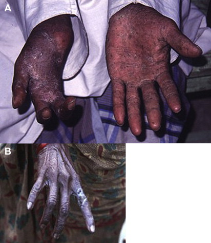
Cutaneous malignancies resulting from arsenic exposure include in situ and invasive SCC ( Fig. 2 ), basal cell carcinoma (BCC), and less often, Merkel cell carcinoma. These malignancies usually develop 10 to 20 years after the initial manifestation of arsenicosis, often as large keratotic nodules. In contrast to the typical sun-exposed distribution of most nonmelanomatous skin cancers, those resulting from arsenic exposure tend to occur in nonexposed sites. In addition, the malignancies may be multiple and can arise both in areas of existing keratoses and in uninvolved skin. Invasive SCC that develops within keratoses in patients with a history of arsenic exposure may be aggressive with a heightened metastatic risk. BCCs arising in the setting of arsenic exposure tend to be multiple and may resemble in situ SCC clinically.
Risk Factors
Several studies have indicated that men are more susceptible to the arsenic-related skin effects than women, a finding that may be explained by metabolism. A population-based study in Bangladesh showed a strong association between more efficient methylation of arsenic and decreased risk of developing skin lesions in women as compared with men. Additional risk factors for skin lesions include tobacco use, sun exposure, increasing age, folate deficiency, hyperhomocysteinemia, and low urinary creatinine excretion. Genetic variability affecting the metabolic capacity of arsenic also contributes to risk for skin lesions.
Laboratory Findings
Arsenic exposure can be established by testing environmental sources, such as the water being consumed, and by monitoring the patient. Biologic monitoring as a measure of arsenic exposure most commonly includes measuring arsenic concentration in urine, hair, nail, and serum. Urine is the primary route of elimination of most arsenic species. Spot urine samples are taken commonly, being easy and painless to collect. Previous data have suggested that the level of arsenic in urine does not vary significantly over time and can be used as a long-term biomarker of arsenic exposure provided that the exposure is current and ongoing. Urine samples taken more than 24 to 48 hours after the last exposure can underestimate peak exposure because most of the arsenic would have been excreted. In addition, the subject should not have consumed any seafood for 4 days before urine collection. Hair and nail samples can be useful to estimate the average amount and rate of arsenic exposure within the previous 9 months. However, these samples can be influenced by external contamination by environmental arsenic exposure. According to the WHO 2005 field guide for the detection of arsenicosis, a urine sample with arsenic concentration greater than 50 μg/L confirms recent exposure. Dry hair with a concentration greater than 1 mg/kg or nail with a concentration greater than 1.5 mg/kg may indicate of exposure to unsafe doses of arsenic within the previous 11 months. Serum concentrations are not so commonly used to screen for chronic exposure because arsenic is rapidly cleared from the blood; serum testing is most useful to assess recent high-concentration exposures.
Histologically, premalignant and malignant, cutaneous, arsenic-related tumors are generally indistinguishable from their counterparts due to ultraviolet radiation. Together with clinical and social history, the absence of significant dermal solar elastosis may be a helpful clue to identify the cause. Arsenical keratoses typically demonstrate compact parakeratosis overlying an acanthotic epidermis with mild keratinocytic dysplasia. There may also be some basal keratinocyte vacuolization and a chronic inflammatory dermal infiltrate. As in non-arsenic–related SCC in situ, full-thickness intraepidermal atypia with parakeratotic hyperkeratosis, without invasion of the dermis, characterizes arsenic-related SCC in situ. When BCC is caused by arsenic, it is usually of the superficial pattern; however, nodular, reticulated, and pigmented patterns of BCC may all be associated with chronic arsenic exposure. Arsenic-related BCCs may demonstrate vacuolated cells, dyskeratosis, multinucleated giant cells, and increased numbers of atypical nuclei and mitotic figures.
Management and Prevention
Management and prevention of the cutaneous sequelae of chronic arsenic exposure is multifaceted. Symptomatic treatment of arsenical keratoses with or without pigmentary changes includes the use of topical 5% to 10% salicylic acid and 10% to 20% urea ointment. Treatment of arsenic-related cutaneous neoplasms includes surgical excision, cryosurgery, electrodesiccation and curettage, oral retinoid therapy, and topical chemotherapy. Imiquimod 5% cream used once daily for 6 weeks has been found effective against arsenical keratoses, SCCs, and BCCs. Dietary modifications and vitamin supplementation to influence the methylation and subsequent detoxification of arsenic have been found to reduce the deleterious effects of arsenic on the skin. These measures include supplementation with vitamin E, selenium, folic acid, riboflavin, and pyridoxine, with the effects of the B vitamins possibly being additive. The levels at which such nutrients need to be consumed to have a beneficial effect are greater than the current recommended daily amounts. Studies have also confirmed that a lower body mass index is associated with a higher prevalence of skin lesions, supporting that overall malnutrition may increase the risk for arsenic-related skin disease. Chelation therapy with agents such as dimercaptosuccinic acid, dimercaptopropane succinate, and d -penicillamine has variable benefits for patients with chronic arsenicosis.
Cessation of consuming contaminated water is of utmost importance. Arsenic mitigation efforts are underway to address this pressing issue. Interventions that have been implemented include person-to-person reporting of well test results, well labeling, village and individual health education, and installation of more deeply situated wells; in Bangladesh, at least 350-m deep aquifers have been found to be safe from arsenic contamination. In one study, urinary arsenic levels decreased by 46% in people who switched to a well identified as safe. Other proposals to decrease consumption of arsenic-contaminated water include rainwater harvesting, filtration and removal of arsenic from current water supplies via individual devices or treatment plants, and treatment of surface water with pressure filtration and disinfection.
Arsenical keratoses and their sequelae are significant contributors to morbidity resulting from chronic exposure to arsenic in drinking water in Bangladesh and they may also serve as indicators for further systemic evaluation for disease. It is to be hoped that a combination of mitigation proposals and programs with surveillance by the medical community for latent sequelae and public health education and counseling will mitigate the deadly impact of chronic arsenic exposure in this population and guide future efforts worldwide.
The authors have nothing to disclose.
Related posts:
 Buruli Ulcer: Advances in Understanding Mycobacterium ulceransInfection
 Outbreak of Nontuberculous Mycobacterial Disease in the Central Pacific
 Chagas Disease: Coming to a Place Near You
Dermatology in Botswana: The American Academy of Dermatology’s Resident International Grant
Chagas Disease: Coming to a Place Near You
Dermatology in Botswana: The American Academy of Dermatology’s Resident International Grant
 Widespread Use of Toxic Skin Lightening Compounds: Medical and Psychosocial Aspects
Human Immunodeficiency Virus and Leprosy: An Update
Widespread Use of Toxic Skin Lightening Compounds: Medical and Psychosocial Aspects
Human Immunodeficiency Virus and Leprosy: An Update
Stay updated, free articles. Join our Telegram channel

Full access? Get Clinical Tree


