Infantile hemangioma is the most common soft tissue tumor of childhood. Despite its frequency, it has only been in the last decade that these lesions have been better characterized and become the subject of significant clinical and translational research. Although most infantile hemangiomas are uncomplicated and do not require intervention, they can be a significant source of parental distress, cosmetic disfigurement, and morbidity. The wide spectrum of disease in the morphology of these lesions and in their behavior has made it difficult to predict the need for treatment and has made it challenging to establish a standardized approach to management.
Key points
- •
Infantile hemangioma is the most common soft tissue tumor of childhood.
- •
Although spontaneous resolution of these lesions occurs, they can be a significant source of functional impairment and present risk for permanent cosmetic disfigurement.
- •
Awareness of risk factors predictive of complications and the need for treatment are key to ensuring appropriate management and optimizing patient outcomes.
- •
Discovery of the efficacy of β-blockers for the treatment of IH and its perceived lower side effect profile than medications traditionally used have resulted in widespread use. As the widespread use in infants is relatively new, the nature and true frequency of adverse effects are still being determined, and recommendations for monitoring are expected to evolve.
Infantile hemangioma (IH), or hemangioma of infancy, is the most common soft tissue tumor of childhood. Despite their frequency, it has only been in the last decade that these lesions have been better characterized and have become the subject of significant clinical and translational research. Recognition of the importance of defining subtypes and risks of IH has begun to lay the groundwork to develop future studies to create evidence-based guidelines for management and to care for affected patients better. Although most IHs are uncomplicated and do not require intervention, they can be a significant source of parental distress, cosmetic disfigurement, and morbidity. The wide spectrum of disease both in the morphology of these lesions and, more importantly, in their behavior has made it difficult to predict the need for treatment and has made it challenging to establish a standardized approach to management.
Epidemiology
The incidence of IHs has been difficult to ascertain because IHs may not appear until after the immediate newborn period; as such, they have not traditionally been included in birth defect registries or surveillance systems. Inconsistent nomenclature used in older studies evaluating the incidence of hemangioma, whereby terms included “strawberry mark,” “cavernous hemangiomata,” “hemangioma,” “strawberry nevi,” and “angiomas,” has made the interpretation of the findings less reliable. A recent prospective study of newborns followed for the first 9 months of life demonstrated an incidence of 4.5% overall, with close to 10% in preterm infants. Demographic risk factors for the development of IH include Caucasian race, female gender (female-to-male ratio of 1.8–2.4:1), and prematurity. Additional perinatal characteristics associated with a higher risk of IH include pre-eclampsia, multiple gestation, and low birth weight. Although some of these observed risk factors are potentially confounding, low birth weight has been found to be the most significant risk factor through multivariate analysis. IHs have previously been considered sporadic; however, clinicians have noted a familial tendency, often caring for multiple siblings with hemangiomas. A recent case-control study in a Chinese population observed that 37% of patients with IH had a history of an IH in a first-degree relative, consistent with an earlier report. Walter and colleagues studied 5 families (22 individuals) with hemangiomas and vascular malformations and found a linkage to a locus on chromosome 5q31–33, suggesting that genes are located on this part of the chromosome, which contributes to the development of hemangiomas. Although these data provide compelling evidence that genetic factors contribute significantly to the development of hemangiomas, to the authors’ knowledge, none of these studies have yet led to identification of a specific gene.
Pathogenesis
Despite the frequency of this tumor, understanding of the pathogenetic mechanisms of IH is still in its infancy. IH pathogenesis is generally believed to be a complex interaction of both genetic and environmental factors. Although early research focused on angiogenesis as the mechanism by which IHs form, more recent efforts have identified postnatal vasculogenesis as a possible pathway for development of IH. Tissue ischemia resulting in neovascularization has been proposed as the stimulus leading to the development of IH. Clinically, in support of this, an area of pallor or decreased blood flow on the skin has been noted to precede the development of IH.
Histologic and immunohistochemical studies have demonstrated that IH is not a neoplasm of normal cutaneous capillaries. Histologically, IHs have markedly increased cellularity with clusters of plump cells that are positive for markers of immature endothelial cells. North and colleagues were the first to note that the endothelial-like cells of the hemangioma expressed GLUT-1, the erythrocyte-type glucose transporter protein that has been shown to be upregulated in zones of hypoxia. GLUT-1 seems to be an exclusive marker for IH and is an invaluable tool used to distinguish hemangiomas from other vascular lesions. The immunohistochemical phenotype of endothelial cells in hemangiomas is identical to that of the chorionic villus cells of the placenta, expressing GLUT-1 at all stages of development, from precursor lesions to fully involuted hemangiomas. Although some have speculated that IH may represent embolization of placenta, an alternative explanation is that the phenotypic similarities instead represent an early, immature progenitor cell, which via its immature fetal phenotype shares common attributes.
In further support of hypoxia’s role in pathogenesis, proliferating hemangiomas have been shown to contain cell types known to preferentially home to hypoxic sites. Hypoxia-induced factors produced by hypoxic endothelial cells play an important role in trafficking of progenitor cells to ischemic tissue. Kleinman and colleagues have demonstrated these factors to be upregulated in the blood (vascular endothelial growth factor A [VEGF-A], matrix metallopeptidase-9) and hemangioma tissue (stromal cell-derived factor-1α, matrix metallopeptidase-9, VEGF-A, and hypoxia-inducible growth factor-1α) from children with proliferating hemangiomas. Recently, Khan and colleagues have characterized multipotential stem cells derived from IH specimens (HemSCs) and have demonstrated their ability to recapitulate human IH in immunodeficient mice, creating the first in vivo model. These hemangioma stem cells and cord blood endothelial progenitor cells are similar to each other in several in vitro assays, suggesting that circulating progenitor cells may be recruited into proliferating IH lesions. The concept that IHs originating from circulating multipotent progenitor cells could also help explain the shared features with placental blood vessels. However, the origin of these hemangioma stem cells remains unknown and remains an area of future study.
Molecular and cellular mediators implicated in the proliferative and involutive phases of hemangiomas include VEGF, basic fibroblast growth factor, insulin-like growth factor-2, tissue inhibitor of metalloproteinase type 1, type IV collagenase, urokinase, hypoxia-inducible growth factor, and mast cells. The importance of the VEGF signaling pathway and its known role in angiogenesis has been identified as a key part of IH proliferation and may account, in part, for the response to corticosteroids and propranolol. Significant investigation into IH pathogenesis is currently underway, which will continue to advance the understanding of the mechanisms involved in IH growth and involution and lead to new, targeted therapy.
Diagnosis
The diagnosis of an IH is typically made clinically based on its appearance and characteristic behavior. Although their appearance may be quite variable, ranging from small, red lesions to large and bulky tumors, it is their behavior that helps to distinguish them from other vascular lesions. Early on, they can appear as a telangiectatic patch or an area of pallor and are often unrecognized until a few weeks of age when they begin to proliferate. The natural history of IH is characterized by an initial proliferative or growth phase followed by a plateau phase, and finally, the involution phase. In the proliferative phase, IHs tend to be firm and noncompressible and become softer and more compressible as they begin to involute. A change in color from bright red to purple or gray can often signal transition to the involution phase. However, the transition from the growth phase to involution may be more dynamic than previously thought, reflecting a balance between local proliferative factors and factors involved in apoptosis. Chang and colleagues detailed the growth characteristics of 526 IHs in 433 children, noting that most hemangioma growth occurs in the first 5 months. However, some IHs exhibit minimal proliferation, remain flat, and may be reticular or networklike in appearance. On average, IHs typically reach their maximum size by 9 months, but deep hemangiomas may proliferate longer. Albeit rare, IHs with an extended growth phase (up to 2 years) have been reported; these tend to be larger lesions and more often segmental or indeterminate rather than localized. A subset of 23 IHs from a large prospective study of 1530 IHs that demonstrated prolonged growth were all of the deep or combined subtype, and it was the deep component that was subjectively thought to have the continued growth in the majority of IHs.
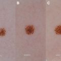
In the past, IHs have been classified by their depth of soft tissue involvement (superficial, deep, and mixed). Superficial hemangiomas ( Fig. 1 ) appear as bright red lesions, which may be plaquelike or more rounded papules or nodules. Deep hemangiomas ( Fig. 2 ) involve the deep dermis and subcutis and present as bluish to skin-colored nodules. Mixed hemangiomas ( Fig. 3 ) clinically have features of both superficial and deep hemangiomas, often with a red plaque overlying a bluish nodule. However, another classification based on morphology has proven to have greater prognostic value and be more predictive of risk of complications or need for treatment. In this classification system, hemangiomas are defined as localized or segmental or indeterminate. Localized hemangiomas are discrete, usually oval or round, and appear to grow from a single focal point. In contrast, the term “segmental” has been used to describe hemangiomas that demonstrate a geographic shape and involve a broad anatomic region or a recognized developmental unit ( Fig. 4 ). The distinction between these morphologic subtypes is an important one, as segmental hemangiomas are at higher risk of complications and associated anomalies. Although the classification of an IH as segmental may be difficult at times to make with certainty, their larger size may assist with recognition of this subtype because they have been shown to cover 4 times greater surface area than localized lesions.
IHs may occur anywhere on the skin, but are most common on the head and neck. Reproducible patterns of segmental hemangiomas on the face have been demonstrated and mapped. Although involvement of the lower face corresponds to known embryologic facial prominences (maxillary, mandibular, and frontonasal), involvement of the upper face (forehead) does not.
Complications
Although most IHs are uncomplicated and do not require treatment, 24% of those referred to tertiary institutions had complications. Providers should be aware of risk factors predictive of complications and the need for treatment to facilitate early referral to a physician with expertise in the management of IHs. Size, location, and subtype (localized vs segmental) are major factors to consider in evaluating an infant’s risk. Specifically, for every 10 cm 2 increase in size, a 5% increase in likelihood of complications as well as a 4% increase in likelihood of treatment has been reported. Although segmental hemangiomas tend to be larger lesions, this subtype has been shown to be an independent risk factor for the development of complications and/or need for treatment. In a prospective IH study reported by Haggstrom and colleagues, segmental IHs were 8 times more likely to receive treatment and 11 times more likely to develop complications compared with localized IHs after controlling for IH size. In this same study, the perceived risk of permanent disfigurement was the leading indication for treatment. The risk of disfigurement depends on the hemangioma subtype, location, and extent of proliferation. Minimally elevated IHs with a gentle slope noted at the edges are at risk for minimal residual telangiectasia or textural change, in contrast to more exophytic IHs with a steeper slope and more prominent superficial component, which are more likely to leave fibrofatty tissue and a resultant scar ( Fig. 5 ). Permanent distortion of anatomic landmarks can occur as the result of location (eg, nasal tip, involvement of ear cartilage) or secondary to ulceration (eg, lip). Additional complications of IHs include ulceration, functional impairment (visual compromise, airway obstruction, auditory canal obstruction, feeding difficulty), and cardiac compromise. Box 1 outlines locations at high risk for the development of hemangioma-specific complications or associated anomalies.
| Location | Potential Complications or Associated Anomalies |
|---|---|
| Periocular | Functional compromise |
| Nasal tip | Cosmetic disfigurement |
| Lip or perioral | Ulceration Cosmetic disfigurement Functional compromise |
| Ear | Ulceration Cosmetic disfigurement Functional compromise |
| Other facial | Cosmetic disfigurement Risk for PHACE syndrome (with segmental morphology) Risk of airway hemangioma (with “beard” distribution) |
| Perineal, perianal | Ulceration Risk of LUMBAR syndrome (with segmental morphology) |
| Lumbosacral | Risk of spinal dysraphism, intraspinal IH, LUMBAR syndrome |
| Hepatic (large) | Congestive heart failure |
Ulceration
Ulceration is the most common complication (reported in 16% of patients in a large prospective study) and can result in pain, infection, bleeding, and permanent scarring. Associated pain can interfere with sleep as well as feeding. Locations at high risk for ulceration and the associated frequency of this complication include anogenital (50%) ( Fig. 6 ), lower lip (30%), and neck (25%). IHs that are larger in size or of the segmental subtype are more likely to develop ulceration. Of the clinical subtypes (ie, superficial, mixed, and deep), the mixed subtype (having both superficial and deep components) has most frequently been associated with ulceration and is another independent risk factor. The cause of ulceration is not well understood, but maceration and friction are likely contributing factors given the higher frequency in locations prone to this. Although ulceration can be complicated by bleeding, clinically significant bleeding (ie, requiring hospitalization/transfusion) is rare.
Visual Compromise
Threat to vision is a common reason for treatment in the IH population. Infants are at particular risk because stimulus deprivation for as little as 1 week can interrupt visual development and result in permanent visual impairment. Periocular IH may cause ptosis, strabismus, and anisometropia, each of which may result in astigmatism, amblyopia, or blindness. In addition, periocular IH can cause proptosis, exposure keratopathy, tear duct obstruction, and rarely, compressive optic neuropathy. Amblyopia can develop in 3 ways, all of which are applicable to periocular IH: refractive-error differences, visual deprivation, and strabismus. Astigmatism, resulting from distortion of the cornea from pressure of the IH, is the most common cause of amblyobia. If the IH affects growth of the eye, anisometropic (refractive error difference) amblyopia can occur. Strabismus may occur in rare cases from direct effects on extraocular muscles. Given the threat of permanent visual impairment, patients with periorbital hemangiomas should be referred early to a physician with an expertise in the treatment of IH and should be closely monitored by ophthalmology, including a retinal examination.
Visceral Involvement and Complications
Although solitary lesions are most common, multifocal cutaneous hemangiomas ( Fig. 7 ) may occur in 30% of patients, although only 3% have greater than 6 lesions. Historically, patients with numerous lesions have been placed into at least 2 categories: disseminated neonatal hemangiomatosis and benign neonatal hemangiomatosis, with the former considered to be more severe with multiple sites of potential extracutaneous disease and a rate of mortality as high as 60%. However, in the past, all multifocal vascular lesions were considered to be hemangiomas, and with advances in histopathologic and radiologic diagnosis (ie, GLUT-1 stain), it is recognized that some of these severe cases represent other multifocal vascular anomalies (ie, multifocal lymphangioendotheliomatosis or cutaneovisceral angiomatosis) rather than true IHs. Many of these other multifocal vascular lesions have a more aggressive course, often with coagulopathy and bleeding, and account for the high mortality historically reported with disseminated neonatal hemangiomatosis. In a critical review of 73 cases reported in the literature as diffuse neonatal hemangiomatosis, 43 were reclassified as IHs or probable IHs with death occurring in only 2 cases (5% of these patients). In contrast, 11 of the 17 (65%) patients were reclassified as multifocal lymphangioendotheliomatosis with thrombocytopenia (MLT)/probable MLT. With greater recognition of other multifocal vascular tumors and improved diagnostics, overly aggressive intervention in infants with asymptomatic multifocal IH may be avoided in the future.
Patients with true multifocal cutaneous IH are recognized to have a higher risk of visceral hemangiomas, with liver and gastrointestinal involvement being most common. Ultrasound of the liver has been recommended in those patients with greater than 5 cutaneous hemangiomas. A recent prospective study investigated the incidence of hepatic involvement in patients with more than 5 cutaneous IHs compared with those with 1 to 4 cutaneous lesions and demonstrated a significantly increased risk in patients with greater than 5 cutaneous lesions. In this study, 24 (16%) of the infants with 5 or more cutaneous IHs had hepatic hemangiomas, whereas none of the infants with less than 5 cutaneous IHs had hepatic hemangiomas ( P <.003), substantiating the recommendation for liver ultrasound in patients with greater than 5 cutaneous IHs.
Similar to cutaneous IH, the nomenclature surrounding liver involvement with IH (infantile hepatic hemangiomas) has been confusing; the term hemangioma has been used to describe venous malformations and IHs in the liver have been referred to as hemangioendothelioma, not to be confused with epithelioid hemangioendothelioma, a malignant tumor with metastatic potential. Infantile hepatic hemangiomas have been subtyped into the 3 following subtypes: focal, multifocal, and diffuse. Focal involvement is typically a solitary lesion that undergoes spontaneous regression and has been likened to the equivalent of a cutaneous rapidly involuting congenital hemangioma; skin involvement in this setting is unusual. Similar to congenital hemangioma and unlike true IH, these lesions are GLUT-1-negative. Multifocal involvement is the subtype typically seen in the setting of multiple cutaneous IHs. Although these are often asymptomatic, high-output cardiac failure may be seen secondary to arteriovenous or portovenous shunting. In a prospective study of infants with multiple cutaneous IHs reported by Horii and colleagues, only 2 of the 24 infants (8%) found to have hepatic involvement required treatment of their hepatic hemangioma. Finally, diffuse involvement with near-total replacement of hepatic parenchyma may be seen. The resulting mass effect can result in abdominal compartment syndrome and multisystem organ failure. Overproduction of type 3 iodothyronine deiodinase by IH resulting in accelerated inactivation of thyroid hormone can result in acquired hypothyroidism in patients with multifocal or diffuse hepatic involvement. These patients should be screened with a serum TSH to ensure that hypothyroidism is not missed, because mental retardation and cardiac failure can ensue if hypothyroidism not detected.
Anomalies Associated with Anatomic Location of IH
The presence of IH in particular locations can be a marker for underlying or associated anomalies. The “beard” distribution ( Fig. 8 ) of an IH, in which preauricular areas, chin, anterior neck, and lower lip is involved, has been associated with the presence of airway hemangiomas. In 2 retrospective studies, 29% to 63% of patients with large IHs on the lower lip, chin, neck, and preauricular region (beard) had airway involvement. Airway hemangiomas typically present between 6 and 12 weeks of age with biphasic inspiratory and expiratory stridor and retractions. Cough may be associated and may mimic croup. Infants with IH in the beard distribution should be monitored closely for respiratory difficulties and referred to an ear, nose and throat specialist for evaluation. Serial evaluations may be required in young infants because the skin hemangioma may precede the development of symptomatic airway IH.