, Shimin Chang2, Jian Lin3 and Dajiang Song1
(1)
Department of Orthopedic Surgery, Changzheng Hospital Second Military Medical University, Shanghai, China
(2)
Department of Orthopedic Surgery, Yangpu Hospital Tongji University School of Medicine, Shanghai, China
(3)
Department of Microsurgery, Xinhu Hospital Shanghai Jiao Tong University, Shanghai, China
The anterior tibial artery perforator (ATAP) flap is vascularized by perforators of the anterior tibial artery and has been reported by several authors [1–6].
23.1 Vascular Anatomy
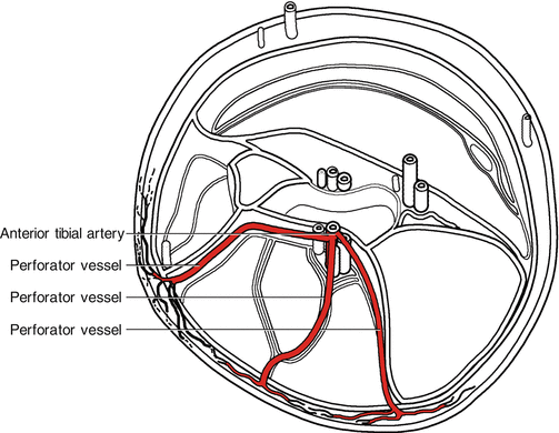
The anterior tibial (AT) artery begins at the inferior border of the popliteus muscle, branching from the bifurcation of the popliteal artery, passes between the two heads of the tibialis posterior, through the interosseous membrane, and to the deep part of the anterior leg. It then descends on the anterior surface of the interosseous membrane, moving closer to the tibia, and becomes the dorsalis pedis artery at the level of the ankle joint. The deep peroneal nerve accompanies the anterior tibial vessels. This is the source of motor branches to the anterior tibial muscle, which branch off from the main trunk within the proximal third of the lower leg (Fig. 23.1).


Fig. 23.1
Vascular anatomy of the anterior tibial artery
Several septocutaneous or musculocutaneous perforators originate from the anterior tibial artery [7, 8].
The most proximal perforators of the anterior tibial artery are approximately 7 cm distal to the fibular head. The perforator then descends through the anterior intermuscular septum between the extensor digitorum longus and peroneus muscles and penetrates the deep fascia in the middle third of the lower leg.
The middle septocutaneous or musculocutaneous perforators are derived from the anterior tibial artery approximately 12 cm distal to the tibial condyle. The perforators course downward through the intermuscular space between the anterior tibial muscle and the extensor digitorum longus muscle to penetrate the deep fascia at the junction of the upper and middle third of the lower leg.
The most distal septocutaneous perforator arises from the anterior tibial system approximately 17–22 cm distal to the tibial condyle and runs caudally beneath the extensor digitorum longus muscle. The vessel courses anteriorly through the lateral intermuscular septum between the extensor digitorum and peroneus muscles or through the medial intermuscular septum between the anterior tibial muscle and the tibia. This perforator finally pierces the deep fascia to enter the skin of the lower two-thirds of the lower leg.
The dual venous system of the ATAP flap comprises the deep venae comitantes of the anterior tibial system or the superficial veins of the small saphenous system.
23.2 Illustrative Case
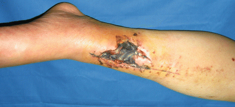
A 35-year-old woman sustained traffic accident. The resulting wound measured 8.5 × 5 cm over the anterior distal third of lower leg (Fig. 23.2).


