Functional reason
Aesthetic reason
Limitations in daily activities
Physical disproportion
Postural alterations
Disability to wear fitted garments
Back pain
Skin rashes in the abdominal fold
The first stage of the abdominal exam is inspection, with the patient standing and in frontal view: assessment of shape, ratio between subcutaneous and deep fat, symmetry, masses, fat distribution, striae, extent and site of dermatochalasis, and shape and position of the umbilicus. Careful attention must be paid to scars, as previous abdominal surgery may have impaired skin vascular supply. Specifically, scar position, length, width, and retraction are accurately analyzed.
The lateral view, in the upright position, allows evaluation of the lateral extent of the abdominal folds (Fig. 13.1). An easy way to assess musculoaponeurotic system laxity is to have the patient bend over (diver’s test) (Fig. 13.2). Standardized photographic documentation is an integral part of the preoperative evaluation [12].
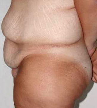
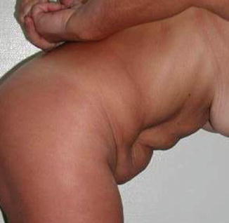
Fig. 13.1
Preoperative
Fig. 13.2
Preoperative diver’s test
The patient is then examined in the supine position for palpation. Guarding, rigidity, or pain is noted and intra-abdominal organs are evaluated. As far as the abdominal wall is concerned, attention is paid to thickness and distribution of the subcutaneous tissue, masses, hernias, and rectus abdominis muscle diastasis. Useful maneuvers to assess hernias are to have the patient cough or hold his/her nose and mouth closed and blow forcibly. Muscle diastasis is better palpated by asking the patient to lift his/her head off the pillow and look at his toes. Skin redundancy is assessed by pinching the abdominal wall horizontally and vertically with the patient standing (Fig. 13.3).
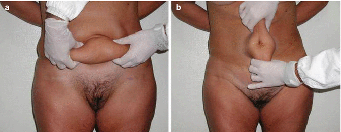
Fig. 13.3
(a, b) Preoperative evaluation of skin redundancy
Percussion is the next step of the abdominal examination and is used to evaluate masses and to determine if abdominal distention is due to gas-filled bowels or accumulation of fluid.
Auscultation for bowel sounds and bruits is the last step of the abdominal clinical examination.
Preoperative laboratory tests are routinely performed when selecting candidates for abdominoplasty, in particular serum proteins and hydroelectrolytic balance must be tested in patients who previously underwent bariatric procedures, especially if malabsorptive. Adequate vitamin and mineral supplementation must be ascertained in order to prevent nutritional deficiencies [13].
ECG and chest X-ray are routine preoperative investigations. A computed tomographic scan or magnetic resonance imaging of the abdominal wall is carried out in all patients with primary or incisional hernias at rest and during straining. Pulmonary function studies are obtained in patients with primary or recurrent abdominal hernias or chronic obstructive pulmonary disease.
This articulate selection allows identification of candidates for abdominoplasty and, based on the specific abdominal wall deformities, the most appropriate surgical approach. Key points are:
1.
Patients who had bariatric surgery with significant abdominal skin excess. In some cases, the excess is mainly evident in the upper abdominal quadrants. In this condition, a conventional abdominoplasty or a body lift would not fully correct the defect.
2.
3.
Advancement of the superior flap, when performing a classical abdominoplasty without excision of the supraumbilical scar, is very limited and leads to unfavorable aesthetic results. In such cases, the median scar resembles a cord under tension.
The anchor-line abdominoplasty offers the opportunity to overcome all these concerns:
1.
Necrosis of the abdominal skin and subcutaneous flaps is unlikely, provided that the lateral segmental vessels are preserved. Dissection is very limited in this procedure and concomitant liposuction to the lateral areas of the abdomen is generally discouraged, to avoid impairment of the vascular supply to the abdominal wall [16, 17]. Liposuction is performed only in selected cases with significant fat deposits in the flanks, and much caution is exercised in the execution of this procedure.
2.
The anchor-line abdominoplasty implies resection of the median or paramedian supraumbilical scars (either vertical or short horizontal) thus preventing their unfavorable stretched appearance.
3.
The upper component of the excision entails a wide area of the abdominal wall, and this is particularly useful in patients with extremely pendulous abdomens. The lateral dermolipectomy allows reduction of waist circumference and improvement of the anteroposterior contour.
It is noteworthy that the vertical and horizontal approach is also advantageous whenever concomitant abdominal or gynecological operations are needed, as it allows easier access to the abdominal cavity. This holds true also in case of recurrent incisional hernias.
13.4 Vascular Anatomy
Huger [18] described the blood supply to the abdominal wall and divided it into three main zones (Huger’s zones I, II, and III) (Fig. 13.4).
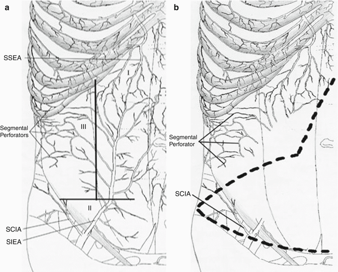
Fig. 13.4
Blood supply to the abdominal wall: (a) Huger’s zones I, II, III (SSEA superficial superior epigastric artery, SCIA superficial circumflex iliac artery, SIEA superficial inferior epigastric artery). (b) The lateral segmental perforators responsible for the vascularization of the flaps advanced in the abdominoplasty
The first zone corresponds to the mid part of the abdomen and is vascularized by the perforating branches of the superior epigastric artery and inferior epigastric artery, which anastomose within the rectus muscle fascia.
The second zone corresponds to the hypogastrium and is nourished by the superficial iliac circumflex artery, the superficial epigastric artery, and some perforating branches from the proximal segment of the inferior epigastric artery. The third zone includes the lateral areas of the abdomen and is supplied by the perforating branches from the diaphragmatic, intercostal, and lumbar arteries.
The third zone is responsible for the vascularization of the lateral cutaneous flaps advanced in the anchor-line abdominoplasty (Fig. 13.4); whenever these lateral segmental branches are spared, there is no increased risk of skin necrosis at the crossing of the vertical and horizontal incisions.
In post-bariatric patients, anatomical variations are observed, as far as the vascular supply is concerned, such as vessel caliber or dislocation.
13.5 Surgical Technique
Bowel preparation with enemas is carried out the day before surgery. Foley catheter is positioned preoperatively. Elastic stockings are applied to the legs to prevent venous stasis, and low-molecular-weight heparin is administered for deep venous thrombosis prophylaxis. Several leading authorities, including The 2006 National Quality Forum (NQF), American College of Chest Physicians (ACCP), and American Society of Colon & Rectal Surgeons (ACRS), endorse the use of prophylaxis against DVT (deep venous thromboembolism) for surgery patients (Table 13.2) [19].
Table 13.2
The 2006 National Quality Forum (NQF), American College of Chest Physicians (ACCP), and American Society of Colon & Rectal Surgeons (ACRS) endorse the use of prophylaxis against DVT (deep venous thromboembolism) for surgery patients
Trends that may reduce DVT risk in general surgery patients: More rapid mobilization Greater use of thromboprophylaxis Other advances in perioperative care Trends that may increase DVT risk in general surgery patients: Performance of more extensive operative procedures in older and sicker patients Use of preoperative chemotherapy Shorter lengths of stay in the hospital, leading to shorter durations of prophylaxis | 2004 American College of Chest Physicians (ACCP) guidelines recommend: Prophylaxis of DVT following abdominal surgerya,b Higher-risk general surgery patients (those undergoing non-major surgery who are >60 years of age or have additional risk factors, or patients undergoing major surgery who are >40 years of age or have additional risk factors): recommend LDUH (5000 U tid) or LMWH (>3400 U daily) (both Grade 1A) High-risk general surgery patients with multiple risk factors: recommend pharmacologic methods (i.e., LDUH tid or LMWH >3400 U daily) combined with the use of GCS and/or IPC (Grade 1C+) Selected high-risk general surgery patients, including those who have undergone major cancer surgery: suggest post-hospital discharge prophylaxis with LMWH (Grade 2A) | 2006 National Quality Forum (NQF)/Surgical Care Improvement Project (SCIP) performance measures outlined for patients undergoing abdominal surgery are: Surgery patients with recommended venous thromboembolism (VTE) prophylaxis ordered Surgery patients who received appropriate VTE prophylaxis within 24 h prior to surgery to 24 h after surgery
Related posts: Abdominoplasty Combined with Cesarean Section: Discussion of the Evidence
Retrospective Analysis of Never Events in Panniculectomy and Abdominoplasty Patients and Their Financial Implications Abdominoplasty Combined with Cesarean Section: Discussion of the Evidence
Retrospective Analysis of Never Events in Panniculectomy and Abdominoplasty Patients and Their Financial Implications
 No Drain Abdominoplasty with Progressive Tension Suture: The Logic, Simplicity, and Aesthetics No Drain Abdominoplasty with Progressive Tension Suture: The Logic, Simplicity, and Aesthetics
 Evolution of the High-Superior-Tension Lipoabdominoplasty: The High-Tension Abdominoplasty Evolution of the High-Superior-Tension Lipoabdominoplasty: The High-Tension Abdominoplasty
 Circumferential Abdominoplasty Circumferential Abdominoplasty
 Abdominal Wall Repair Post Hernia in Kidney and Liver Transplantation Abdominal Wall Repair Post Hernia in Kidney and Liver Transplantation
Stay updated, free articles. Join our Telegram channel
Full access? Get Clinical Tree
 Get Clinical Tree app for offline access

|