AN OVERVIEW OF HAIR FOLLICLE ANATOMY
AND BIOLOGY
By
Paul Mouser Ph.D.
Ashland, Inc.,
Ashland Specialty Ingredients,
Bridgewater, NJ, United States
ABSTRACT
Since ancient times, the human hair has been an attribute for health, youth, and attractiveness as it plays a vital role in people’s self-perception (1). As a result, changes in the appearance of hair such as hair loss, hair thinning, and premature graying often cause psychological distress to the affected individuals.
Hair is produced by a multicellular entity; the hair follicle. The hair follicle is one of the most fascinating and dynamic organs of the human body and, unlike the hair itself, which is composed of dead keratinocytes, hair follicles undergo a process of regeneration that controls the production of the hair fiber. One can distinguish between three main types of hair follicles: the lanugo, the vellus, and the terminal hair follicle:
- ● Lanugo hair is very fine and normally shed in utero, or during the first weeks of life.
- ● Vellus hairs are very short, nonpigmented, nonmedullated hairs found all over the body surface.
Terminal hair is the large, and usually pigmented and medullated hair that is predominantly found on the scalp. It is estimated that humans have around 5 million hair follicles, of which 80,000 to 150,000 are located on the scalp. Scalp hairs grow at a rate of 0.3 to 0.4 mm per day (2). No additional follicles are produced after birth, although the size of the follicles and hairs can alter with time, primarily under the influence of androgens. In pubic, axillary, and beard regions, androgens stimulate the conversion of vellus to terminal hairs, whereas in frontal scalp regions of genetically predisposed individuals, androgens trigger miniaturization of hair follicles via conversion of terminal to vellus hair follicles (3).
The most unique feature of hair growth is its cycle. Hair follicles undergo repeated cycles of anagen (growth), catagen (degeneration), and telogen (quiescence). During anagen, hair follicle stem cells sustain the steady production of matrix cells in the hair bulb. These proliferate several times and terminally differentiate into the companion layer, the inner root sheath, and the hair shaft.
Unwanted hair loss is a common problem and is suffered by much of the world’s population in both women and men. The condition is characterized by premature hair loss and excessive hair shedding. The process is caused by an interruption in the normal hair growth cycle, which can lead to the development of baldness with insufficient replacement of hairs in human scalp. This can have negative effects on an individual’s self-esteem and thus products that claim to be useful for treating hair loss target a steadily growing, multibillion-dollar market worldwide.
The field of human follicle biology is progressing rapidly largely because of technical advances in molecular and cellular biology as well as sophisticated cell culture techniques. This chapter reviews the structure of hair, the biological processes occurring during the hair growth cycle, and the changes that occur with hair aging.
3.3.2.1 Hair Follicle Structure
3.3.2.3 Aging of the Hair Follicle
3.3.2.1 HAIR FOLLICLE STRUCTURE
The hair follicle is a three-dimensional tube, composed mainly of epithelial cells, which protrude down through the epidermis and dermis of the skin (Figure 1).
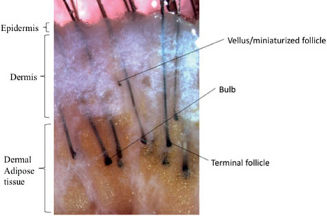
Figure 1. Human scalp tissue isolated from the scalp. Hair follicles protrude down through the epidermis and dermis of the skin (photo kindly provided by Dr Claire Higgins)
Hair follicles vary considerably in shape and size, depending on their location, but they all have common architecture. Anatomically, the hair follicle is most commonly described when it is in the mature anagen stage (Figure 2A). The mature anagen follicle can be divided into an upper permanent portion (infundibulum and isthmus), which does not cycle, and a lower part (bulb and suprabulbar), which is remodeled in each hair cycle. The infundibulum is the opening of the hair canal to the skin surface, and the isthmus is the permanent middle section extending from the sebaceous duct opening to the point of arrector pili insertion. The lower isthmus also contains the bulge region (Figure 2B), which is a region of functional importance as it is in this location that a reservoir of stem cells reside. Stem cells are unique, as they have the capacity to generate all the differentiated cell types within tissue and to self-renew in order to replenish their pool. Earlier studies have demonstrated that all epithelial layers within the hair follicle and hair originated from bulge cells (4, 5). Hair follicle stem cells therefore appear to be responsible for regenerating the hair follicle in each cycle. Up until recently the location of the bulge in human follicles was unclear. By contrast with the bulge in rodent follicles, human follicles are not morphologically distinct. However, identification of unique bulge markers in the human follicle has enabled the human anagen bulge to be defined as a distinct region (6, 7).
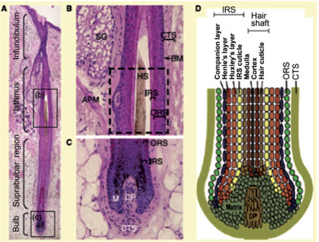
Figure 2. Structure of human anagen hair follicle. (A) Longitudinal section of a human scalp hair follicle showing the permanent (infundibulum and isthmus) and cycling (suprabulbar region and bulb) components of the hair follicle. (B) High magnification image of the isthmus. The dashed square indicates the approximate location of the bulge. (C) High magnification image of the bulb showing the outer root sheath (ORS), inner root sheath (IRS), matrix zone (M), dermal papilla (DP) and connective tissue sheath (CTS). (D) Schematic drawing illustrating the concentric layers of the ORS, IRS and shaft in the bulb. Current Biology 19, R132-R142 2009 ©2009 Elsevier
The anagen bulb (Figure 2C) is one of the most rapidly dividing tissues in the body. The bulb represents the hair shaft factory; it contains the proliferating matrix keratinocytes and pigment-producing melanocytes (matrix zone). The hair matrix keratinocytes move upwards and differentiate into the hair shaft, as well as into the inner root sheath; the melanocytes transfer pigment into the developing hair keratinocytes to give hair its color (8). These cells are supplied by the pluripotent stem cells existing in the bulge. The hair bulb surrounds the dermal papilla, which comprises a group of specialized cells (dermal papilla cells) with important inductive properties. The dermal papilla is believed to be one of the most important drivers that instruct the hair follicle to grow, and dictates hair bulb size, hair shaft diameter, length, and anagen duration.(9) The dermal papilla is known as the “conductor of the orchestra” or “command center,” and is an essential source of growth factors (keratinocyte growth factor, bone morphogenetic protein, hepatocyte growth factor, insulin-like growth factor, stem cell factor), all of which are critical for hair growth and melanogenesis (8). Surgical removal of the dermal papilla and the lower dermal sheath prevents hair growth, which indicates the importance of these specialized cells as the key signaling center in hair follicles (10). Interestingly, dermal papilla cells from balding scalp have both higher levels of androgen receptor (AR) and type II 5-alpha reductase, which converts testosterone to dihydrotestosterone (DHT) (11). DHT can induce dermal papilla cells to secrete factors including transforming growth factor beta 1 (TGFb1) and DKK-1 that inhibit keratinocyte growth (12, 13).
The hair shaft (Figure 2D) is divided into the hair cuticle on the outside and both the cortex and medulla in the center. The cells that make up the hair shaft are known as trichocytes, which are terminally differentiated hair follicle keratinocytes. The hair shaft cuticle consists of overlapping cells that are arranged like shingles pointing outward and upward. The hair shaft cortex, the bulk of the hair shaft, is composed of hair-specific keratin filaments and keratin-associated proteins with melanin produced by melanocytes in the matrix region (2). The hair shaft medulla, the central part of the hair shaft, is composed of large, loosely connected keratinized cells with large intercellular air spaces. The hair shaft is surrounded and supported by the inner root sheath (IRS), the companion layer, and the outer root sheath (ORS).
The IRS consists of three distinct layers: the cuticle, Huxley’s layer, and the Henle’s layer (Figure 2D). The IRS cuticle layer adjoins the cuticle of the hair shaft and anchors the hair shaft to the follicle. IRS keratinocytes produce keratins and trichohyalin that serve as an intracellular “cement” giving strength to the IRS to support and mold the growing hair shaft, as well as guide its upward movement (8).
The ORS surrounds the IRS and is continuous with the basal layer of the epidermis. Distinct from the other epithelial components, the ORS does not generate by upwards growth from the matrix zone, but rather directly from the stem cell bulge. The bulge region of the ORS is located just below the sebaceous gland and at the insertion site of the arrector pili muscle. Stem cells leave the bulge in the ORS and migrate toward the matrix zone, where they begin to proliferate and subsequently differentiate into new hair shafts (14).
Hair follicles grow in an oblique angle towards the epidermal surface. This angle can be varied by the contraction of a bundle of muscle, collectively called the arrector pili muscle (APM). The APM is under adrenergic control and thus, in situations of cold temperature and emotional stressors, it gives rise to the interesting phenomenon of involuntary contraction resulting in the hair standing up!
Related posts:
 – AN OVERVIEW OF THE CHANGING REGULATORY LANDSCAPE IN THE U.S. AND THE E.U. AND HOW TO DEAL WITH THEM…
 3 – THE SUBSTRATES
 – CLASSIFICATION SCALE FOR SKIN COMPLEXIONS AROUND THE WORLD
 – ASIAN ETHNIC SKIN: SPECIALTY CORRECTIVE COSMECEUTICALS FOR ASIAN ETHNIC SKIN CARE
 – THE NOSE: ACCESSING THE BIOLOGY OF HUMAN OLFACTION: NEW, ALL-NATURAL FRAGRANCE INGREDIENTS; NOVEL CONSUMER FRAGRANCE EXPERIENCES AND APPLICATIONS
 – MECHANISMS OF CHANGES IN HAIR SHAPE
Stay updated, free articles. Join our Telegram channel

Full access? Get Clinical Tree


