Clinical criteria
1. Acute cutaneous lupus
Including lupus malar rash (do not count if malar discoid)
Bullous lupus
Toxic epidermal necrolysis variant of SLE
Maculopapular lupus rash
Photosensitive lupus rash in the absence of dermatomyositis
Or subacute cutaneous lupus
Nonindurated psoriasiform and/or annular polycyclic lesions that resolve without scarring, although occasionally with postinflammatory dyspigmentation or telangiectasias
2. Chronic cutaneous lupus
Including classical discoid rash
Localized (above the neck)
Generalized (above and below the neck)
Hypertrophic (verrucous) lupus
Lupus panniculitis (profundus)
Mucosal lupus
Lupus erythematosus tumidus
Chilblain lupus
Discoid lupus/lichen planus overlap
3. Oral ulcers
Palate, buccal, tongue, or nasal ulcers
In the absence of other causes, such as vasculitis, Behcet’s disease, infection (herpes), inflammatory bowel disease, reactive arthritis, and acidic foods
4. Nonscarring alopecia
(Diffused thinning or hair fragility with visible broken hairs)
In the absence of other causes such as alopecia areata, drugs, iron deficiency, and androgenic alopecia
5. Synovitis
Involving two or more joints, characterized by swelling or effusion or tenderness in two or more joints and 30 min or more of morning stiffness
6. Serositis
Typical pleurisy for more than 1 day
Or pleural effusions
Or pleural rub
Typical pericardial pain (pain with recumbency improved by sitting forward) for more than 1 day
Or pericardial effusion
Or pericardial rub
Or pericarditis by EKG
In the absence of other causes, such as infection, uremia, and Dressler’s pericarditis
7. Renal
Urine protein/creatinine (or 24 h urine protein) representing 500 mg of protein/24 h
Or
Red blood cell casts
8. Neurologic
Seizures, psychosis mononeuritis multiplex
In the absence of other known causes such as primary vasculitis
Myelitis, peripheral or cranial neuropathy
In the absence of other known causes such as primary vasculitis, infection, and diabetes mellitus
Acute confusional state
In the absence of other causes, including toxic-metabolic, uremia, drugs
9. Hemolytic anemia
10. Leukopenia or lymphopenia
Leukopenia (<4,000/mm3 at least once)
In the absence of other known causes such as Felty’s syndrome, drugs, and portal hypertension
Or
Lymphopenia (<1,000/mm3 at least once) in the absence of other known causes such as corticosteroids, drugs, and infection
11. Thrombocytopenia
(<100,000/mm3) at least once
In the absence of other known causes such as drugs, portal hypertension, and TTP
Immunological criteria
1. ANA
Positive, above laboratory reference range
2. Anti-dsDNA
Positive, above laboratory reference range, except ELISA: twice above laboratory
Reference range
3. Anti-Sm
Positive
4. Antiphospholipid antibody
Any of the following lupus anticoagulant:
False-positive RPR
Medium- or high-titer anticardiolipin (IgA, IgG, or IgM)
Anti-β2 glycoprotein I (IgA, IgG or IgM)
5. Low complement
Low C3
Low C4
Low CH50
6. Direct Coombs test
Positive, in the absence of hemolytic anemia
The Revised Cutaneous Lupus Erythematosus Disease Area and Severity Index (RCLASI) has specifically been developed to assess disease activity and damage in CLE. This disease activity index focuses on LE skin parameters, including scaling, hypertrophy, dyspigmentation, edema, infiltration, and subcutaneous nodules [1].
9.2 The Spectrum of Cutaneous LE
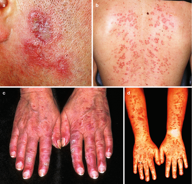
As indicated above, the spectrum of skin lesions which may appear in the context of LE is wide. To make a complex topic even more complicated, these LE-associated skin lesions are distinguished in two major groups: “LE-specific” and “LE-nonspecific” lesions. The LE-specific lesions encompass all the specific dermatological subsets of LE and are clinically subdivided into four different subtypes (acute LE/ACLE, subacute cutaneous LE/SCLE, intermediate cutaneous LE/ICLE, and chronic cutaneous CLE). Typical clinical examples for these CLE subsets are depicted in Fig. 9.1. Skin lesions typically associated with autoimmune diseases albeit not LE-specific are, amongst others, vasculitis, livedo racemosa, calcinosis cutis, and skin ulcers [1].


Fig. 9.1
The clinical heterogeneity of cutaneous lupus erythematosus skin lesions. Chronic discoid LE: (a) discoid erythrosquamous plaques in the face; (b) subacute cutaneous LE: annular maculae at the back; (c) chilblain LE: erythematous maculae and nodules of the hands; (d) bullous LE (vesicles and small bullae in sun-exposed skin)
As detailed in Table 9.2, two complementary strategies can be used to classify the different specific subsets of CLE: (1) a clinical classification, focusing on the different dynamics of the CLE subsets, and (2) a histological classification, which is based on the typical histological picture and the anatomical structures involved. The typical clinical and histological findings of the most common CLE subtypes are summarized in Table 9.3.
Table 9.2
Clinical and histological classification of CLE
Clinical classification of CLE modified from Kuhn and Landmann [1] | |
CLE | |
Acute CLE (ACLE) | Localized form |
Generalized form | |
Bullous LE | |
Subacute CLE (SCLE) | Annular form |
Papulosquamous form | |
Intermediate CLE (ICLE) | Lupus erythematosus tumidus (LET) |
Chronic CLE (CCLE) | Discoid lupus erythematosus (DLE) |
Localized form | |
Disseminated form | |
Lupus erythematosus profundus (LEP; LE panniculitis) | |
Chilblain lupus erythematosus (CHLE) | |
Histological classification of CLE: | |
Histological pattern | LE subtype |
Dermoepidermal LE | Acute LE |
Subacute LE | |
Chronic discoid LE | |
Bullous LE | |
Chilblain LE | |
Dermal LE | LE tumidus |
Jessner’s lymphocytic infiltration (JLI) | |
Reticular erythematous mucinosis (REM) | |
Papular mucinosis | |
Hypodermal LE | LE panniculitis (LE profundus) |
Table 9.3
Clinical and histological findings of the most common CLE subtypes
Clinical | Histological | |
|---|---|---|
Acute CLE (ACLE) | Butterfly rash | Edema and only mild lesional inflammation with a cell-poor interface dermatitis |
Erythema in sun-exposed skin | Neutrophils within the infiltrate | |
Close association to SLE | ||
ANA >90 % positive | ||
Anti-dsDNA, anti-RNP, and anti-Sm often positive | ||
Subacute cutaneous LE (SCLE) | Annular, gyrated, or plaque-like erythrosquamous lesions in sun-exposed skin, but not the face | Cell-poor IFD with superficial lymphoid infiltrate |
ANA 60–80 % positive | Atrophic epidermal layer | |
Anti-SSA/Ro and anti-SSB/La often positive | Mucin deposits in upper dermis | |
LE tumidus (LET) | Erythematous, pad-like, infiltrated lesions in sun-exposed skin (including the face) | Dense, patch-like, perivascular, and perifollicular lymphoid infiltrate (superficial and deep) |
No scaling | Large amounts of mucin within the dermal layer | |
ANA 10–30 % positive | Clusters of CD123-positive pDCs | |
Anti-SSA/Ro and anti-SSB/La negative | ||
Chronic discoid LE (CDLE) | Scarring, discoid, and erythrosquamous lesions | Dense, patch-like, perivascular, and perifollicular lymphoid infiltrate (superficial and deep) |
Predilection sites: capillitium and face | Cell-rich IFD, involvement of the follicular epithelium and follicular hyperkeratosis | |
Positive tin-tack sign | Broadened basement membrane | |
ANA 10–30 % positive | Lots of mucin within the dermis | |
LE profundus (LEP) | Early: subcutaneous nodules and indurations | Early: lobular panniculitis with numerous lymphocytes and pDCs |
Late: fat-tissue atrophy and skin retraction | Late: necrosis, fibrosis, and macrophages | |
Predilection sites: gluteal region, thigh, upper arms, and face | Mucin deposits in the subcutis | |
ANA ~75 % positive | Polyclonal TCR rearrangement (DD: cytotoxic T-cell lymphoma) | |
Chilblain LE (ChLE) | Livid and painful with erythemata and indurations and nodules of fingers and toes | Dense, perivascular lymphoid infiltrates within the dermis |
Special subset: familial ChLE with mutation on TREX1 gene | Fibrin deposits within small dermal vessels | |
Sometimes additional typical findings of other CLE subsets (IFD, mucin) | ||
Bullous LE | Small vesicles on erythematous skin in sun-exposed skin areas | Subepidermal blisters with neutrophils |
Close association to SLE | Direct immunofluorescence: linear deposits of IgG, IgM, IgA, and C3 | |
Anti-collagen VII positive |
9.2.1 Acute CLE (ACLE)
Acute courses of CLE clinically present with plane redness in sun-exposed skin areas, particularly in the face, décolleté, and extensor sides of the arms. The most characteristic clinical sign is the butterfly rash, a symmetric, circumscribed, aliform erythema on both cheeks. This CLE subset shows a close association to SLE in younger female patients and may be accompanied by fever, myalgia, fatigue, and involvement of internal organ systems. In the peripheral blood, often high-titer antinuclear antibodies with SLE-typical specificity (e.g., anti-dsDNA, anti-Sm) are detectable. In contrast to the impressive clinical picture, the histological changes may be discrete. The slides show a mild edema with only minor mucin deposits. However, some neutrophils may accompany the dermal and junctional inflammatory infiltrate. These are rarely found in other CLE subsets and may be indicative for ACLE [5].
9.2.2 Bullous CLE (BLE)
The BLE is a very rare subset of ACLE which presents clinically with confluating small vesicles on erythematous skin in sun-exposed skin areas. Circulating anti-collagen VII antibodies, which target structures in the basement membrane, are typically found in the peripheral blood of the patients affected. Histologically, skin lesions show a subepidermal blister (effect of the anti-collagen VII antibodies) and a neutrophil-rich inflammatory infiltrate (close association to ACLE/SLE).
9.2.3 Subacute Cutaneous LE (SCLE)
The SCLE presents clinically with widespread annular, gyrated, and/or plaque-like erythrosquamous lesions in sun-exposed skin areas including décolleté and extensor sides of the arms, while the face typically is not involved. The majority of SCLE patients are ANA-positive. Among the ANA, anti-SSA/Ro and anti-SSB/La antibodies are the most characteristic antibodies for this CLE subset. These antibodies not only are important diagnostic markers but also have a functional impact, since newborn babies of anti-SSA/Ro-positive mothers have an increased risk to develop SCLE-like skin lesions which diminish with the decline of the maternal postpartal immunoglobulin protection. From a histological point of view, SCLE shows a cell-poor interface dermatitis with vacuolar degeneration of the basal epidermal layer, colloid bodies, immigration of skin-homing lymphocytes, and dermal mucin deposits [6, 7].
9.2.4 LE Tumidus (LET)
LET clinically presents with infiltrated erythematous maculae and plaques without epidermal scaling which primarily appear in sun-exposed skin areas including the face. LET shows a dynamic which is between subacute CLE and chronic CLE and therefore was classified as “intermediate CLE type” in the Düsseldorf classification of CLE [1]. Histologically, the lesions are characterized by an extensive dermal spot-like perivascular and perifollicular inflammatory lymphoid infiltrate without any epidermal component. This inflammation is dominated by CD123-positive plasmacytoid dendritic cells, which form clusters of more than ten cells together. This inflammation is accompanied by strong dermal mucin deposits. Most of the patients are ANA-negative, and SLE-typical organ manifestations are uncommon in LET. Particularly, anti-SSA/Ro and SSB/La, which are frequently found in other photosensitive CLE subsets, are almost completely lacking [8, 9].
9.2.5 Chronic Discoid LE (CDLE)
The most common CLE subset is the chronic discoid LE (CDLE). It presents clinically with chronic, discoid, scarring, erythrosquamous plaques particularly affecting the capillitium and face. Most manifestations remain localized to one body region, but extensive courses with widespread scarring lesions appear in up to 10 % of the cases. In contrast to the extensive lesional skin damage, CDLE patients have only a minor risk to develop SLE, and the ANA titers are usually low. The most typical clinical sign of CDLE is the so-called tin-tack sign: a painful hyperkeratosis with follicular plugging due to the follicular hyperkeratosis of CDLE. Histologically, the lesions show a dense periadnexal and perivascular lymphoid inflammation, accompanied by a cell-rich interface dermatitis with a broadened basement membrane. Older lesions are characterized by fibrosis, due to the scarring character of this CLE subset [5].
9.2.6 LE Profundus (LEP)
In early stages, patients suffering from LEP show subcutaneous nodules and pad-like indurations of the dermis and subcutis. During the course of the disease, the patients develop lipoatrophy with retractions of the skin. The gluteal region is the predilection site of the disease, but the thighs, upper arms, and the face may also be involved. Histologically, LEP (also called “lupus panniculitis”) shows a lobular panniculitis with several CD8-positive cytotoxic T cells, accompanied by CD123-positive plasmacytoid dendritic cells and lesional expression of type I IFN-regulated cytokines. CLE-typical changes may also be seen in the dermis (e.g., mucin depositions) and at the dermoepidermal junction (interface dermatitis) in individual cases [10–12].
9.2.7 Chilblain LE (ChLE)
Stay updated, free articles. Join our Telegram channel

Full access? Get Clinical Tree


