Trichoscopy facilitates the diagnosis of various hair and scalp disorders and is often useful in predicting the disease course. However, to date, few studies describe the dermoscopic findings unique to Afro-textured hair. This article reviews what is currently known regarding trichoscopy and discusses its usefulness in this population.
Key points
- •
Trichoscopy of Afro-textured hair and scalp is still evolving.
- •
Pinpoint white dots are a unique dermoscopic feature of the scalp in people of African ancestry and make diagnosis of scarring alopecia more difficult.
- •
Dermoscopy is useful in selecting the biopsy site and, ultimately, in increasing the rate of pathologic diagnosis.
Introduction
Dermatoscopy is a noninvasive technique that has gained wide use in “in vivo” diagnosis of hair diseases. Hair dermatoscopy, also referred to as trichoscopy, facilitates visualization and analysis of scalp and hair structures and patterns. These structures include hair shafts, follicular openings, cutaneous vascular patterns, and perifollicular signs. Given the variation in reproducibility of current diagnostic standards (eg, clinical examination, pull test, and biopsy), trichoscopy serves to improve diagnostic accuracy. The extensive study and use in the white population has made it an integral technique used in daily dermatologic practice.
However, to date, little has been published regarding the dermatoscopic findings in hair disorders of Afro-textured hair. Although many of the same diseases affect all hair types, the distinct properties of the hair and scalp in patients of African descent warrant further investigation into the trichoscopic patterns unique to this population. For example, the hair shaft in African American patients is tightly curled in tertiary structure. In cross-section, it is flattened or elliptical. It is common to observe knots and areas of breakage along the hair shaft, which can be easily visualized with the dermatoscope ( Fig. 1 ). The hair follicles are similarly curved. Less moisture is also characteristic of hair shafts in African American patients compared with whites. Moreover, the scalp is often more darkly pigmented. This article reviews the current knowledge regarding dermatoscopic findings specific to Afro-textured hair and suggests when trichoscopy-guided biopsy may be appropriate in affected individuals.
Normal scalp
Scalp color in dark-skinned individuals can vary from light brown to dark black on dermatoscopy but does not necessarily correlate with the actual color of the skin. Another typical feature is a perifollicular pigmented network or honeycomb pattern. The presence of rete ridge melanocytes account for the hyperchromic lines of this pattern, whereas the hypochromic areas have fewer melanocytes residing in the suprapapillary epidermis. Hair density is significantly lower than in white patients (about 18 terminal and 3 vellus hairs in a 4 mm punch) but the hair diameter is larger.
Each follicle contains one or two vellus hairs and between two and four terminal hairs. In general, the hairs display a coarser, curlier texture. A characteristic feature of the pigmented scalp is the presence of small white dots, which have been named pinpoint white dots. Pinpoint white dots appear as small, 0.2 to 0.3 mm, white dots distributed regularly between the follicles among the pigment network ( Fig. 2 ). These white dots were originally described by Kossard and Zagarella in the scalp a patient with lichen planopilaris (LPP), but they were misinterpreted as a sign of fibrosis. Abraham and colleagues coined the term pinpoint white dots. They correlated this dermoscopic feature with the opening of the sweat ducts. More recently, reflectance confocal microscopy showed that the pinpoint white dots correspond to acrosyringeal and follicular openings. Pinpoint white dots make distinguishing scarring from nonscarring alopecia more difficult than in the nonpigmented scalp because the loss of follicular openings is not immediately evident. Scaling is often a prominent feature, possibly related to the high prevalence of seborrheic dermatitis in this population. Vessels are often visible but there are no studies describing the vascular patterns of the normal black scalp.
Tinea capitis
Tinea capitis (TC) is a common dermatophyte infection affecting children, particularly those of African descent. The most commonly reported dermatoscopic findings associated with TC in black children are corkscrew hairs. These were first described by Hughes and colleagues in African children with endothrix TC, particularly of the Trichophyton soudanense type. Since then, corkscrew hairs have also been found in TC caused by T violaceum . In fact, Vazquez-Lopez and colleagues documented the resolution of corkscrew hairs in TC of an African American child following administration of griseofulvin therapy. These broken, twisted hairs are usually seen in association with comma hairs, which are typically seen in TC of black and white patients ( Fig. 3 ). Comma hairs, originally described by Slowinska and colleagues in patients with Microsporum canis infection are broken, comma-shaped hairs that can easily be distinguished from the blunt-ended, tapering hair shaft, exclamation mark hairs in alopecia areata. The comma shape is thought to result from hair shaft cracking and bending because of ectothrix or endothrix fungal infection. Corkscrew hairs are exclusive to African descent patients, in whom they are seen in endothrix and ectothrix infections. Broken hairs and black dots are also observed. The cause for corkscrew hairs is unknown; however, it is likely linked to the anatomic shape of African hairs. Furthermore, the coiled hairs have been found to have many more fungal elements compared to the straight hairs.
Alopecia areata
Alopecia areata is a common, nonscarring, hair loss disorder. It occurs in whites and African Americans with equal frequency. Despite its prevalence, however, only one study has described the dermatoscopic findings seen in an African American patient. As in normal scalp, the honeycomb-like network can be seen in alopecic patches. Yellow dots are usually absent and the alopecic area shows an increased number of pinpoint white dots. Diagnosis is based on presence of exclamation mark hairs, broken hairs, and black dots ( Fig. 4 ).
Alopecia due to hair breakage
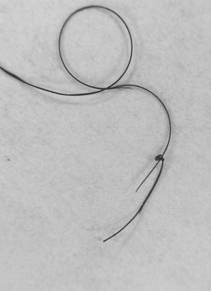
Hair breakage is common because of the intrinsic characteristics of African hair and the hair shaft damage caused by multiple styling processes. Severe hair breakage, predominantly caused by chemical procedures can cause alopecic patches. Patients complain that their hair does not grow and present a positive tug sign. Dermoscopy is helpful to show breakage with Trichorrhexis nodosa , which can be seen in the scalp or, even more easily, in the shed broken hairs ( Fig. 5 ).
Related posts:
 The Evolution of Skin Pigmentation and Hair Texture in People of African Ancestry
What’s New in Objective Assessment and Treatment of Facial Hyperpigmentation?
Cosmeceuticals
Folliculitis Keloidalis Nuchae and Pseudofolliculitis Barbae
The Evolution of Skin Pigmentation and Hair Texture in People of African Ancestry
What’s New in Objective Assessment and Treatment of Facial Hyperpigmentation?
Cosmeceuticals
Folliculitis Keloidalis Nuchae and Pseudofolliculitis Barbae
 New Insights on Keloids, Hypertrophic Scars, and Striae
Clinical Presentations of Severe Cutaneous Drug Reactions in HIV-Infected Africans
New Insights on Keloids, Hypertrophic Scars, and Striae
Clinical Presentations of Severe Cutaneous Drug Reactions in HIV-Infected Africans
Stay updated, free articles. Join our Telegram channel

Full access? Get Clinical Tree


