Fig. 1
Pre-operative anteroposterior view radiograph obtained at initial presentation. Note the large intermetatarsal angle
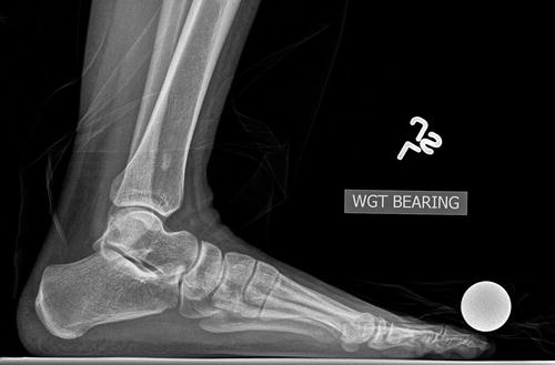
Fig. 2
Pre-operative lateral view radiograph of the foot at initial presentation. Note the slight elevation of the first metatarsal
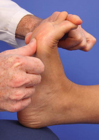
Fig. 3
Pre-operative lateral view clinical photograph of the foot assessing hypermobility of the first ray at initial presentation. Note the left thumb is loading the lateral column of the foot to mimic the mid stance phase of gait and the index finger is loading the hallux to mimic propulsion phase of gait. Hypermobility is clinically present when the first metatarsal head can be elevated during the exam with the opposite thumb. This patient did demonstrate hypermobility of the first ray
3 Preoperative Problem List
1.
Bunion
2.
Large first intermetatarsal angle
3.
Hypermobility of the first ray
4 Treatment Strategy
Distinct patient advantages exist with external fixation including the ability to undergo bilateral foot surgery, immediate weight bearing, participation in showering or pool rehabilitation, the option of returning to work earlier, caring for family, independent living, and no retained internal fixation after treatment completion. Weight bearing begins immediately after surgery with a wooden bottom surgical shoe. The external fixation construct may be removed once radiographic osseous healing is identified. The patient’s foot is protected for 1 month after the external fixation removal by using a surgical shoe while weight bearing as tolerated. Patients are instructed to perform first metatarsal phalangeal joint range of motion exercises at home or with prescribed physical therapy sessions, once the skin incisions are healed, both after the initial surgery and upon the removal of external fixation.
5 Basic Principles
External fixation is placed remote from the incisional approach for the Lapidus procedure to avoid a pin site infection creating incisional infection.
It provides the surgeon a dynamic control of the perioperative process and the ability to make effective clinical decisions which positively impact the surgical outcome.
The external fixation includes at least two points of orthogonal fixation per osseous segment.
Immediate weight bearing serves to increase regional blood flow to the bone and reduces complications associated with prolonged non-weight bearing such as deep vein thrombosis, pulmonary embolism, osteopenia, deconditioning/muscle atrophy, cast disease, and potential for obesity from inactivity.
External fixation has the ability to adjust to what radiographic or clinical changes occur by either adding compression or dynamization to the external fixation which has positive effect on bone healing.
6 Images During Treatment
See Figs. 4, 5, 6, 7, and 8.
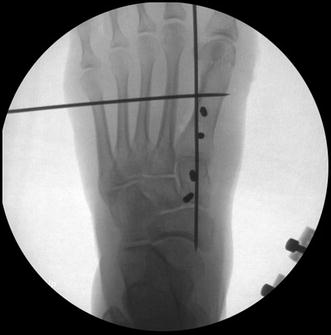
Fig. 4
 8: Femoral Bone Defect
 11: Bone Transport Over a Nail for Infected Tibial Nonunion and Bone Defect
 22: Bone Transport to a Knee Fusion and Secondary Intramedullary Nailing s/p Gunshot Wound
 39: Ilizarov Ankle Fusion
 63: Residual Clubfoot: Equinovarus Deformity/Knee Valgus/Limb Length Discrepancy
 65: Closed Correction of Club Foot with Ilizarov


Related posts:
8: Femoral Bone Defect
11: Bone Transport Over a Nail for Infected Tibial Nonunion and Bone Defect
22: Bone Transport to a Knee Fusion and Secondary Intramedullary Nailing s/p Gunshot Wound
39: Ilizarov Ankle Fusion
63: Residual Clubfoot: Equinovarus Deformity/Knee Valgus/Limb Length Discrepancy
65: Closed Correction of Club Foot with Ilizarov
Stay updated, free articles. Join our Telegram channel
Full access? Get Clinical Tree
