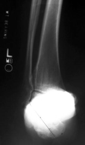
Fig. 1
Talar declination 10° (17 contralateral) and calcaneal pitch 20° (26 contralateral)

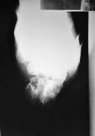
Fig. 2
Calcaneal deformity compared to contralateral normal side: 40° varus, 25 mm short, 23 mm loss of hindfoot height

Fig. 3
Calcaneal bone appearance consistent with previous infection and multiple surgical procedures

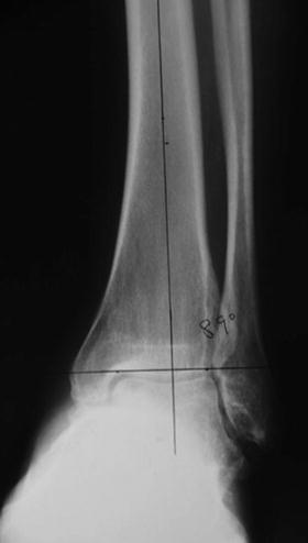
Fig. 4
LDTA 89°
3 Preoperative Problem List
1.
Calcaneal varus and shortening
2.
Calcaneal bone defect with history of osteomyelitis
3.
Compromised soft tissue envelope due to infection and multiple prior procedures
4.
Loss of employment
4 Treatment Strategy
The treatment strategy comprised of gradual calcaneal deformity correction with lengthening and realignment. A medial hindfoot incision was used (avoiding the compromised lateral soft tissues). The calcaneal osteotomy was made via multiple drill holes adjacent to the previously infected osteotomy site. A Taylor spatial frame was applied in a butt frame construct. Gradual correction of deformity was performed, correcting hindfoot varus, restoring calcaneal length, and improving limb length. The frame was modified, placing a standard foot ring after deformity correction to facilitate weight bearing and function.
5 Basic Principles
Pre-operatively, deformity analysis involves ankle, foot, and hindfoot alignment view radiographs. Gradual correction without internal fixation was implemented for severe deformity, shortening, risk of neurovascular injury, compromised soft tissues, and prior infection. Frame construct was optimized for ease of correction and then modified to facilitate weight bearing and function.


