Fig. 1
Medial side view of the right foot
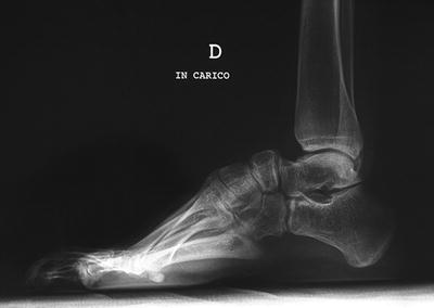
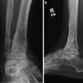
Fig. 2
Lateral radiograph of the right foot before surgery
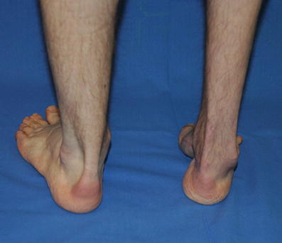
Fig. 3
Posterior view of the both feet
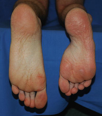
Fig. 4
Plantar view of both feet demonstrates the length discrepancy
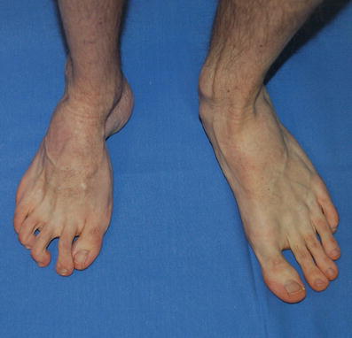
Fig. 5
Dorsal view of both feet takes in evidence of adduction of the forefoot
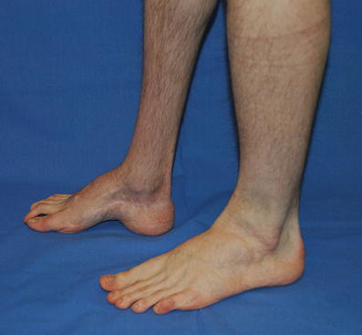
Fig. 6
Clinical lateral view of both feet before treatment
3 Preoperative Problem List
(a)
Varus deformity of the calcaneus
(b)
Cavus deformity of the midfoot
(c)
Adduction and supination of the forefoot
(d)
Shortening of all feet
4 Treatment Strategy
The pre-operative plan included osteotomy of the posterior calcaneus for correction of varus and osteotomy at the level of the cuboid and navicular bone for correction of cavus deformity and adduction and supination of the forefoot.
5 Basic Principles
(a)
Gradual separate correction of the calcaneus with progressive lengthening
(b)
Gradual separate correction of the midfoot in the level of osteotomy and fixation of the fragments of the cuboid and navicular with cutted olive wire for more guided correction of the part of the midfoot
(c)
Fixation of the talus to maintain the relationship in the ankle joint
6 Images During Treatment
See Figs. 7, 8, 9, 10, 11, 12, 13, and 14
 8: Femoral Bone Defect
 11: Bone Transport Over a Nail for Infected Tibial Nonunion and Bone Defect
 22: Bone Transport to a Knee Fusion and Secondary Intramedullary Nailing s/p Gunshot Wound
 39: Ilizarov Ankle Fusion
 63: Residual Clubfoot: Equinovarus Deformity/Knee Valgus/Limb Length Discrepancy
 65: Closed Correction of Club Foot with Ilizarov


Related posts:
8: Femoral Bone Defect
11: Bone Transport Over a Nail for Infected Tibial Nonunion and Bone Defect
22: Bone Transport to a Knee Fusion and Secondary Intramedullary Nailing s/p Gunshot Wound
39: Ilizarov Ankle Fusion
63: Residual Clubfoot: Equinovarus Deformity/Knee Valgus/Limb Length Discrepancy
65: Closed Correction of Club Foot with Ilizarov
Stay updated, free articles. Join our Telegram channel
Full access? Get Clinical Tree
